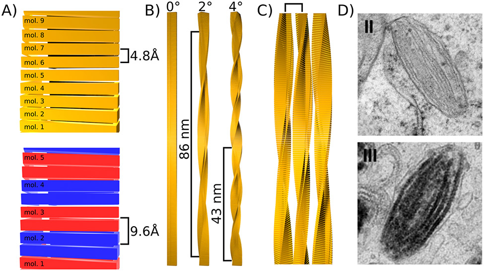
Figure 2: Levels of periodicity in cross-β fibrils.
A) Periodicity along the fibril axis caused by the repetition of monomers. In the simplest case when every strand of a β-sheet is formed by a different monomer, the periodicity is 4.8 Å (top). Otherwise the this periodicity is multiple of 4.8 Å. For example when one monomer provides two strands of the same β-sheet as illustrated with alternating blue and red monomers (bottom). B) Periodicity along the fibril axis caused by a slight rotation of each monomer around the fibril axis (yaw). Three examples with rotation angles of 0°, 2°, and 4° are given and the resulting periodicity is indicated. C) Periodicity induced by lateral association of fibrils. A square bracket indicates the resulting periodicity. D) Example of lateral association of Pmel17 fibrils during stage II of melanosome formation (top), to which melanin binds during stage III (bottom). Images from Hurbain and co-workers (2008) Copyright 2008 National Academy of Sciences.

