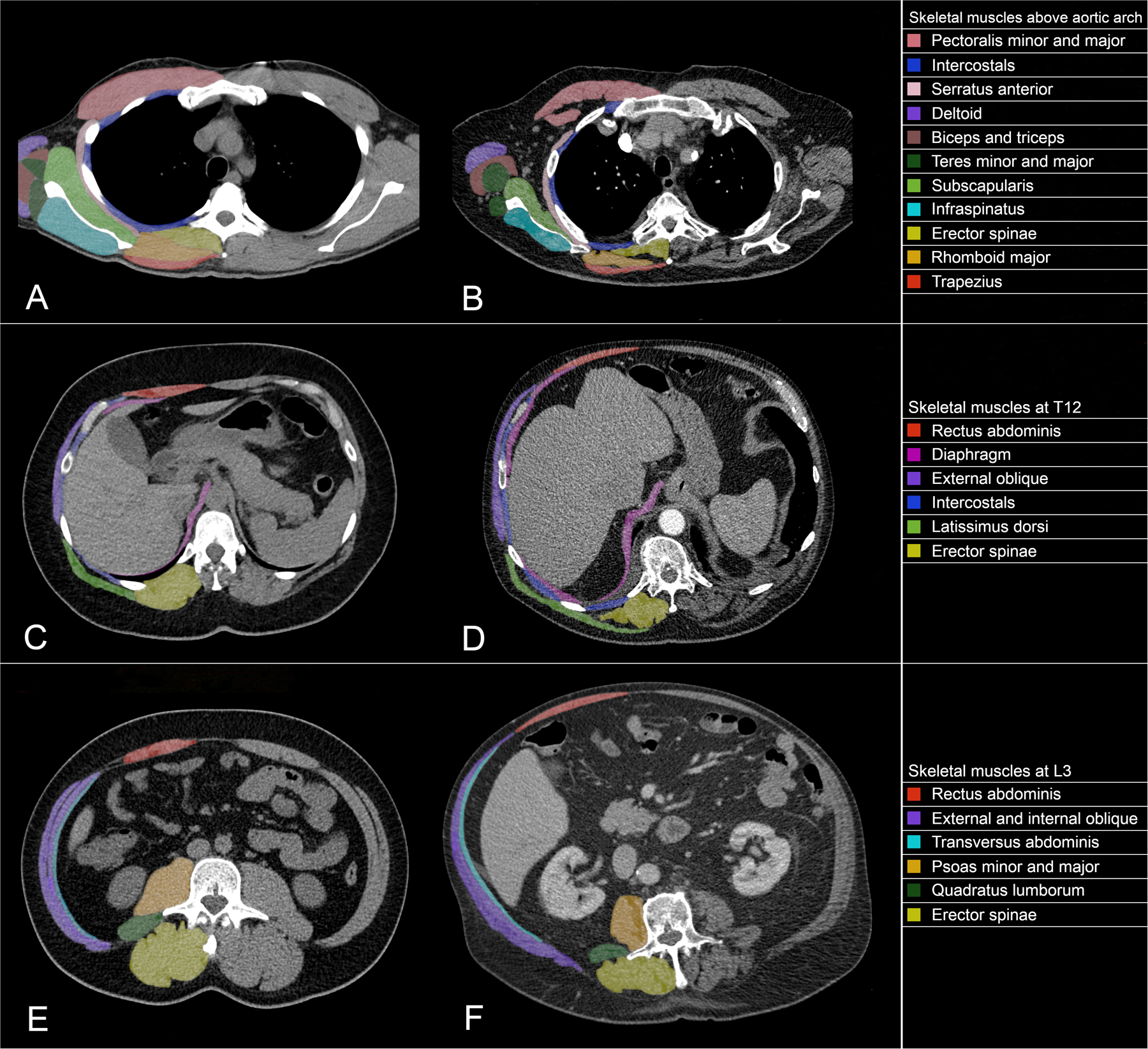
Figure 3.

Computed tomography axial slices demonstrating the skeletal muscles found at the most commonly studied vertebral levels, including normal (2A) and low (2B) muscle mass above the aortic arch (about the third thoracic vertebra), normal (2C) and low (2D) muscle mass at the twelfth thoracic vertebra, and normal (2E) and low (2F) muscle mass at the third lumbar vertebra.
