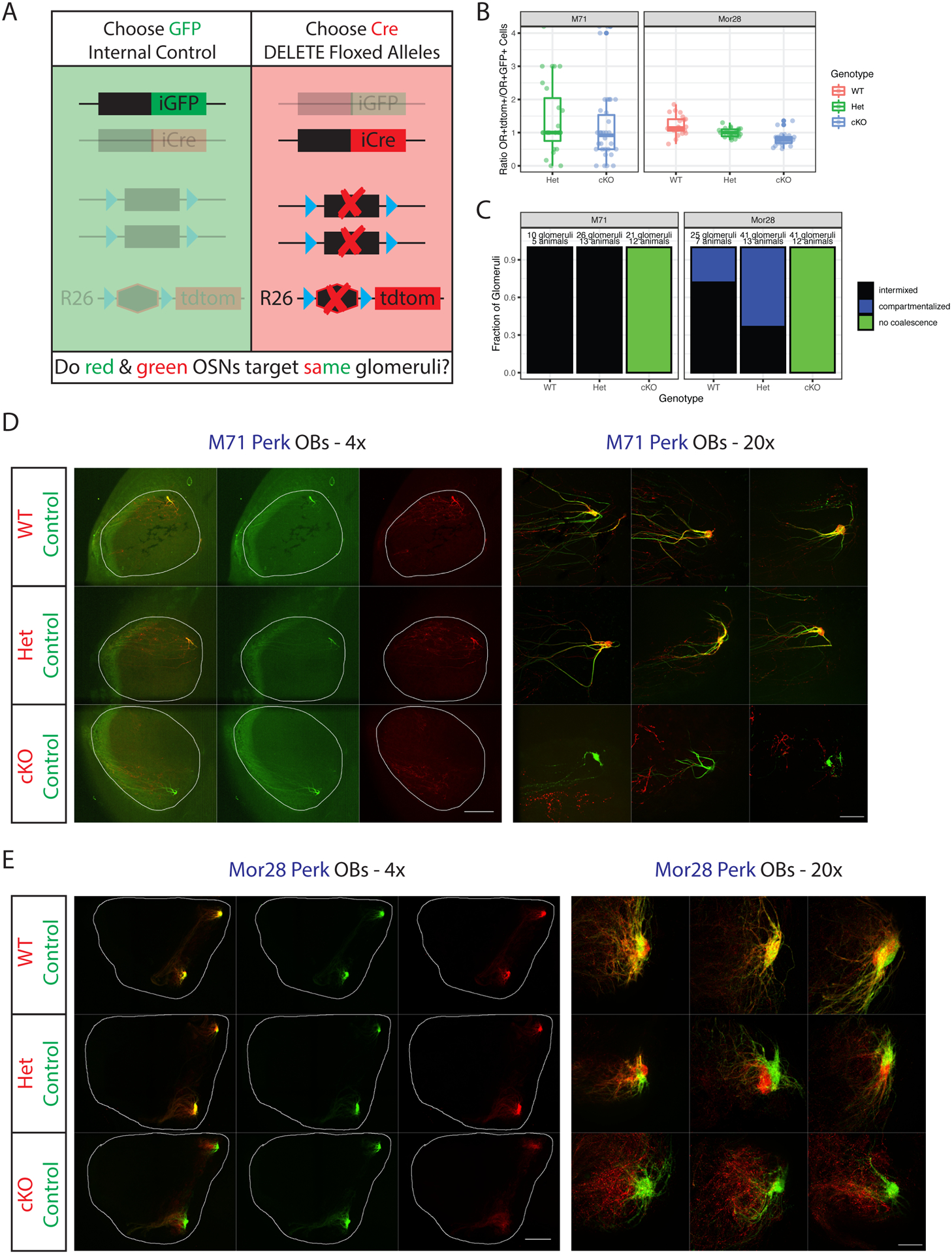
Figure 4. Continuous PERK signaling is required for OSN axon coalescence.

(A) Schematic of the monoallelic deprivation strategy. Mice have the genotype OR(iresCre/iresGFP); Rosa26(LSL-tdtom/+) with or without additional floxed alleles. Targeting of red axons is compared to internal control green axons.
(B) Ratios of OR+tdtomato+ to OR+GFP+ cells in the MOE of p5 mice. Data facetted by OR type and colored by genotype of the tdtomato+ cells. Each point is a section, n=3 mice per genotype except for M71 Het (where n=2). P>0.05 for all comparisons except Mor28 cKO-WT (p=0.0038) by one-way ANOVA with Tukey’s post-test. Also see Supplemental Figure S4A–B.
(C) Blinded quantification of glomerular configurations in the OB of p5 M71 and Mor28 Perk mice, grouped by genotype of the tdtomato+ cells. Also see Supplemental Figure S4D–E, Supplemental Table S4.
(D) Whole mount OB views of M71 Perk mice at p5. Internal control axons are green, experimental axons red with the indicated genotypes. Magnification as indicated, A=anterior, P = posterior. Scale bars 500μm (4x), 100μm (20x).
(E) Same as D but for Mor28 Perk. Also see Supplemental Figure S4C.
