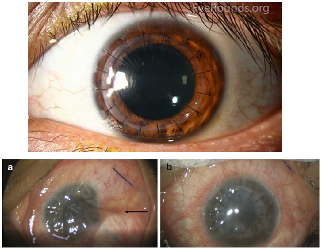
Figure 1: Penetrating Keratoplasty:

Top, grafts for penetrating keratoplasty with 16 interrupted sutures. Reproduced with permission from the Department of Ophthalmology at the University of Iowa, and available at EyeRounds.org. Accessed 24March22. Below are intra-operative photographs from a different case of dehisced penetrating keratoplasty. (A) Note the deflated globe with indentation of the sclera (black arrow) and corneal edema. (B) After repair, the return of the globe contour and wound closure with ten interrupted sutures can be seen. Reproduced with permission from the publisher from Davies E., Yonekawa Y. (2018) Case 6: Dehiscence of Penetrating Keratoplasty from Blunt Trauma. In: Grob S., Kloek C. (eds) Management of Open Globe Injuries. Springer. https://doi.org/10.1007/978-3-319-72410-2_11
