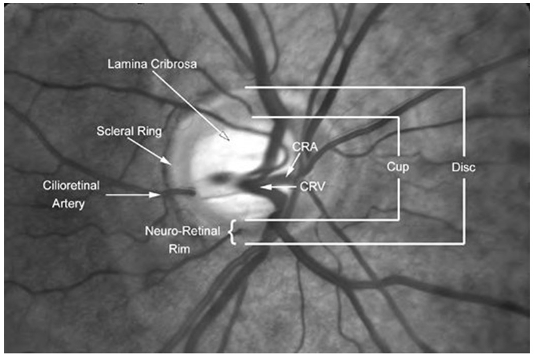
Figure 2A: The components of the cup and disc in the fundus image of the eye.

In a low cup-to-disc ratio, the axons that constitute the optic nerve are at risk of compression as they exit the eye in the lamina cribrosa. In a high cup-to-disc ratio, there is a higher risk of glaucoma. Reproduced with permission, from novel.utah.edu and Dr. Kathleen Digre, the copyright holder. A link to the figure is available at https://collections.lib.utah.edu/ark:/87278/s6d24vxw. Accessed 24March22
