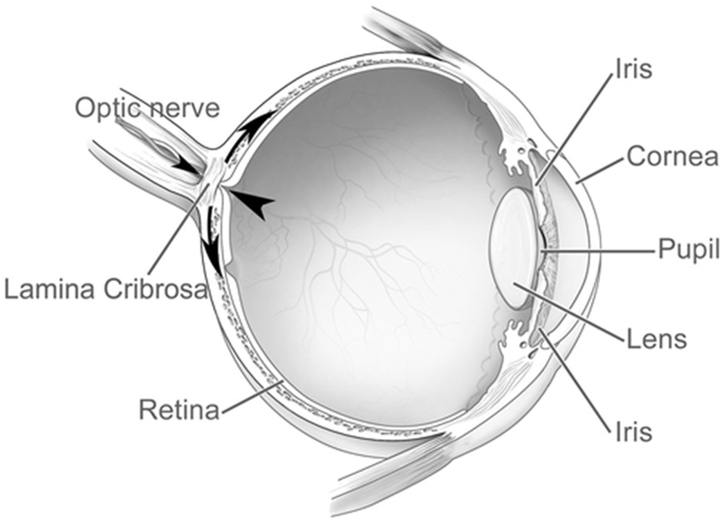
Figure 2B: Cross section of the eye illustrating locations of retina, optic nerve, and the lamina cribrosa.

Large arrowhead shows the pressure exerted by the intraocular pressure, and the small arrowhead the retrobulbar cerebrospinal fluid pressure. The intraocular pressure also generates a pressure load to the inner surface of the eye wall, as shown by the curved arrows. Reprinted with permission from the publisher from Imaging of the lamina cribrosa and its role in glaucoma: a review. Ching-Yu Cheng, Michaël JA Girard, Victor Koh, et al, Clinical & Experimental Ophthalmology, John Wiley and Sons, Jan 10, 2018
