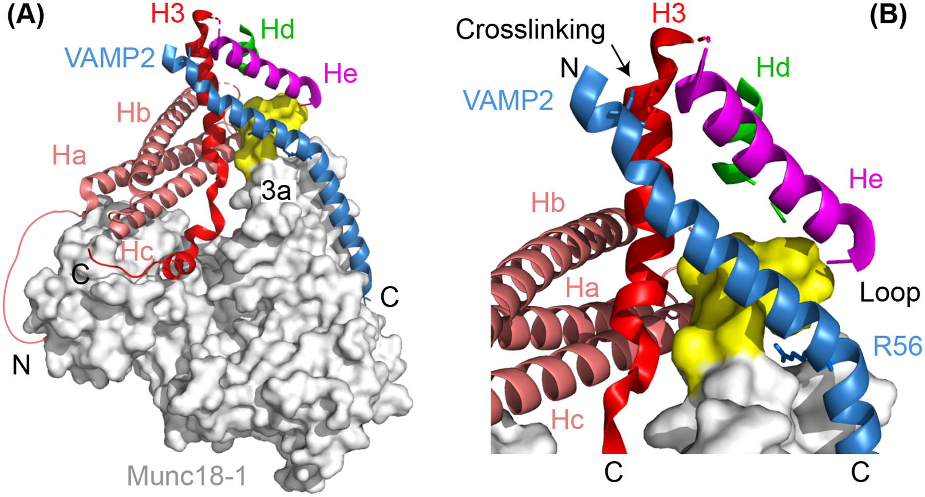
Figure 5.

Atomic structure of the template complex (A) and the close-up view of the N-terminal four-helix bundle (B). In (A), the helical N-peptide binds to the back of Munc18–1 (grey) and connects to the Ha helix with a disordered polypeptide. Part of the unfurred loop of the 3a helical hairpin in Munc18–1 (a.a. 327–334) exhibits a helical conformation and is highlighted yellow, while the other part (a.a. 316–326) is not resolved and likely disordered. The amino (‘N’) - and carboxyl (‘C’) -ends of the SNARE polypeptides are labelled. In (B), the zero-layer residue R56 in VAMP2 and the cysteine residues in both VAMP2 and Syntaxin-1 used for crosslinking the two polypeptides are shown in sticks.
