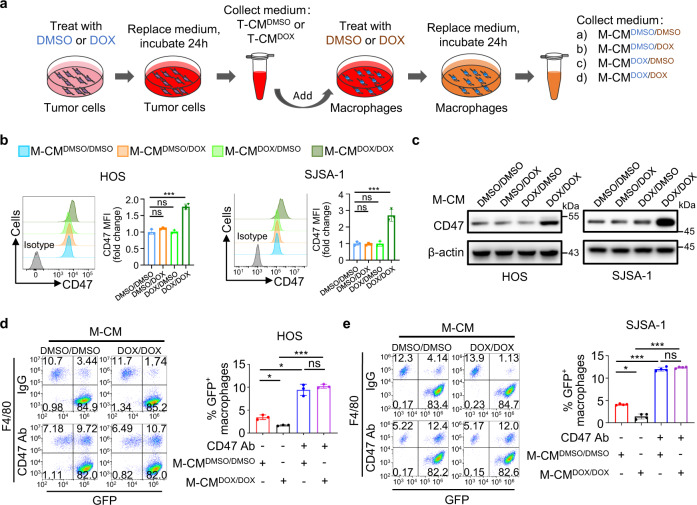
Fig. 3. Macrophage-secreted factors induced by doxorubicin promote CD47 expression in osteosarcoma cells for inhibition of macrophage phagocytosis.
a Schematic diagram of producing macrophage-conditioned media (M-CM). HOS cells were treated with DMSO or doxorubicin (DOX) to produce DMSO-treated tumor-conditioned medium (T-CMDMSO) or DOX-treated tumor-conditioned medium (T-CMDOX). THP-1-cell-derived macrophages were treated with the T-CMDMSO or T-CMDOX in the presence of DMSO or DOX to produce four kinds of M-CM: M-CMDMSO/DMSO was produced after treated with T-CMDMSO and DMSO; M-CMDMSO/DOX was produced after treated with T-CMDMSO and DOX; M-CMDOX/DMSO was produced after treated with T-CMDOX and DMSO; M-CMDOX/DOX was produced after treated with T-CMDOX and DOX. b, c HOS or SJSA-1 cells were treated with M-CMDMSO/DMSO, M-CMDMSO/DOX, M-CMDOX/DMSO, or M-CMDOX/DOX for 24 h. b CD47 expression on the cell surface was determined by flow cytometry using isotype control or anti-CD47 antibody (left). The anti-CD47 median fluorescence intensity (MFI) was determined (right; n = 3 independent experiments). c Western blot analysis of CD47 expression in tumor cells. d, e Flow cytometry-based in vitro macrophage-mediated phagocytosis assay of HOS (d) or SJSA-1 (e) cells in the presence of IgG or anti-CD47 antibody (CD47 Ab) for 4 h (n = 3 independent experiments for HOS cells and n = 4 independent experiments for SJSA-1 cells). HOS or SJSA-1 cells were pretreated with M-CMDMSO/DMSO or M-CMDOX/DOX for 24 h. Macrophages were defined as F4/80+ (labeled with PE) events, and tumor cells as GFP+. F4/80+, GFP+ events represented macrophages that had phagocytosed tumor cells. Data are shown as the mean ± SD. ns not significant. *P < 0.05, ***P < 0.001, one-way ANOVA (b, d, e). The experiment was performed three times with similar results (c). See the Source Data file for the exact P values. Source data are provided as a Source Data file.

