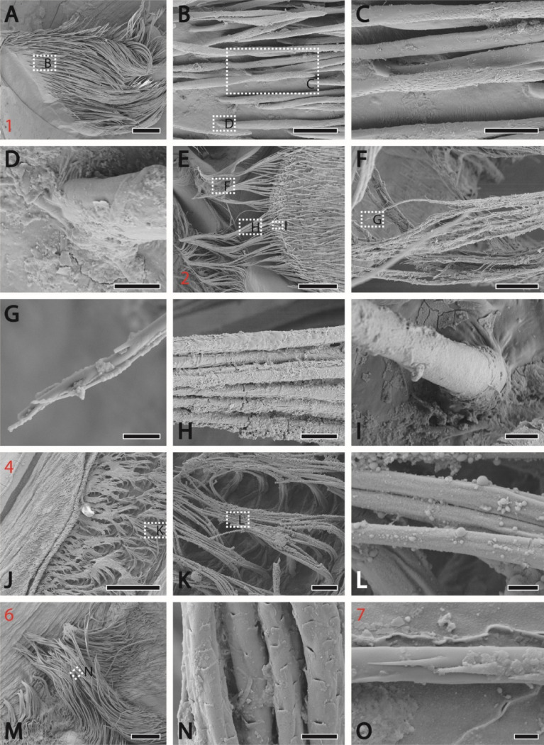
Figure 3.
SEM images of the setae from distinct localities (the locality number is highlighted in red). (A) Presumably cuspidate setae with bold shaft and tip bearing small projections from the locality 1 with magnifications (B)–(D). (D, I) Attachment of the setae with the membrane; no socket could be seen. (E) Setae [also at high magnifications (F)–(I)] bearing long serrated setules (plumodenticulate setae), which agglomerated and formed a net, from the locality 2. (J) From the locality 4, long setae [with magnifications (K)–(L)] without setules of denticles, clustering together forming mattes. (M) From the locality 6; long setae [at high magnification (N)] bearing small scales (pappose setae). (O) Setae with small projections on the tip from the locality 7. Scale bars: A, E, 400 µm; B, 80 µm; C, F, 40 µm; D, 8 µm; E, H, K, 20 µm; F, 100 µm; G, L, 3 µm; I, N, 6 µm; J, 200 µm; K, 20 µm; M, 200 nm; O, 2 µm.

