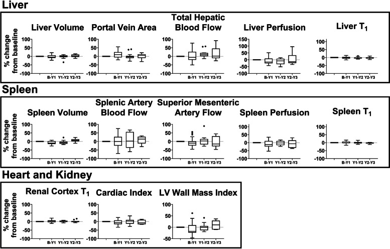
Fig. 4.
Year-to-year percentage change in magnetic resonance imaging measures in the stable compensated cirrhosis control group. Measures liver (volume, portal vein area, total hepatic blood flow, liver perfusion, liver T1), spleen (volume, splenic and superior mesenteric artery flow, spleen perfusion, spleen T1), kidney (renal cortex T1), and heart (cardiac index and left ventricle [LV] wall mass index) are shown. Bars indicate interquartile range and horizontal bold line shows the median percentage change, dots represent outliers

