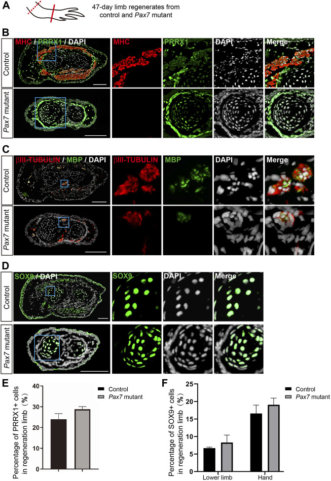
FIGURE 5.
Examination of cell types in limb regenerates in Pax7 mutants. (A) Scheme of the sample collection. Dotted line, amputation plane; line, the position of analyzed section. (B–D) Immunofluorescence images of MHC (red), PRRX1 (green) and DAPI (white) (B); βIII-TUBULIN (red), MBP (green) and DAPI (white) (C); and SOX9 (green) and DAPI (D), in cross-sections, from 47-days fully regenerated control and Pax7 mutant limbs. Boxed regions are shown at higher magnification as separated channels. Scale bars, 200 μm. (E,F) Quantification of the percentage of PRRX1 (E) and SOX9 (F) positive cells, in the limb from controls (n = 3) and Pax7 mutants (n = 3). Data are presented as mean ± sem.

