
FIG. 2.
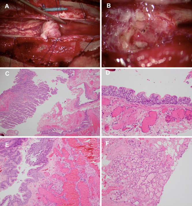
Intraoperative view (A) of the cyst showing both a solid and a cystic component filled with clear, gelatinous fluid. Cerebrospinal fluid flow was reestablished after resection (B). Hematoxylin and eosin stain demonstrating at low power (×4 power) a partially disrupted cyst with a pseudostratified epithelial lining that forms tufts into the cyst lumen (C). Higher magnification (×20 power) shows the cyst lining to have prominent cilia (D). The cyst walls (×10 power) contain prominent dilated and congested capillaries (E) in some areas, as well as serous and mucinous glands with some adipose tissue in others (F).