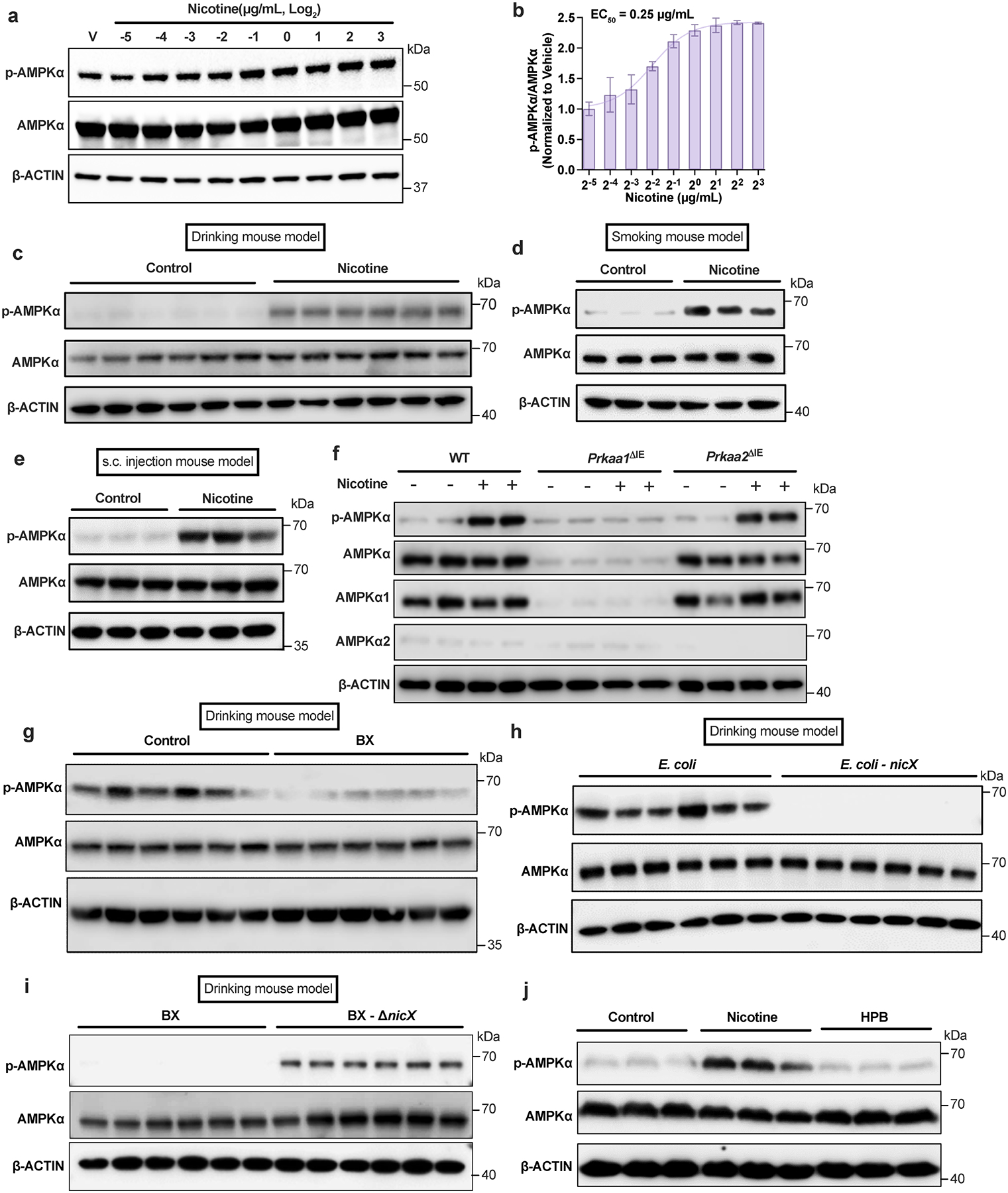
Extended Data Fig. 4. Nicotine-induced activation of intestinal AMPKα.

a, b, Activation of the AMPKα in ileal organoids after treatment with nicotine at different concentrations for 4 h (n = 3 independent experiments). c, Western blot analysis indicated that ileal AMPKα was activated in the nicotine drinking mouse model (SPF, n = 6 mice/group). d, e, Western blot analysis indicated that ileal AMPKα was activated in the smoking mouse model (d) and the subcutaneous injection mouse model (e) (SPF, n = 3 mice/group). f, Western blot analysis of ileal primary enterocytes isolated from WT, Prkaa1ΔIE and Prkaa2ΔIE mice (SPF) and then cultured with or without nicotine (1 μg/mL) treatment for 4 h. Experiments were performed with n = 4 mice/group. g, Western blot analysis showing ileal AMPK signaling in the nicotine drinking mouse model transplanted with Control or B. xylanisolvens (SPF, n = 6 mice/group). h, i, Western blot analysis showing ileal AMPK signaling in the nicotine drinking mouse model transplanted with E. coli or nicX knock-in E. coli (h); and WT or nicX knock-out B. xylanisolvens (i) (SPF, n = 6 mice/group). j, Western blot analysis showing AMPK signaling in SW480 cells incubated with nicotine (1 μg/mL) or HPB (1 μg/mL) for 4 h. This result is representative of 3 independent experiments. In c-e, g-i, mice were supplied nicotine plus HFHCD for two weeks. Data are the means ± s.e.m. (b).
