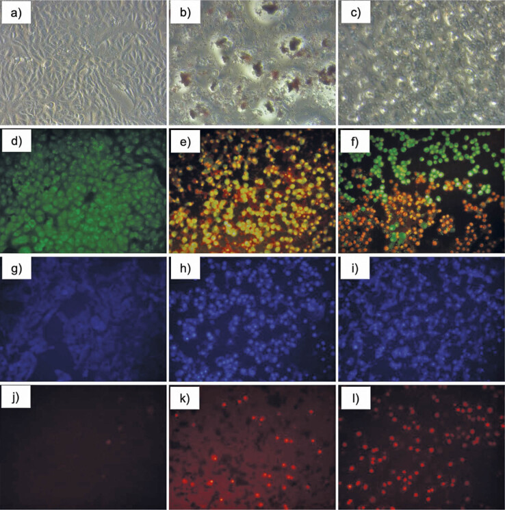
Fig. 3.
Morphology of MDA-MB-231 cells after treatment with IC50 of capsanthin extract or capsanthin-loaded micelles of diosgenin polyethylene glycol succinate 1000 (cap-DPGS-1000): a−c) control and inverted-phase contrast images after treatment with capsanthin extract and cap-DPGS-1000, respectively, d−f) control and AO-EB staining images after treatment with capsanthin extract and cap-DPGS-1000, respectively, g−i) control and Hoest-33342 staining images after treatment with capsanthin extract and cap-DPGS-1000, respectively, j−l) control and propidium iodide staining images after treatment with capsanthin extract and cap-DPGS-1000, respectively. Control=stained untreated MDA-MB-231 cells

