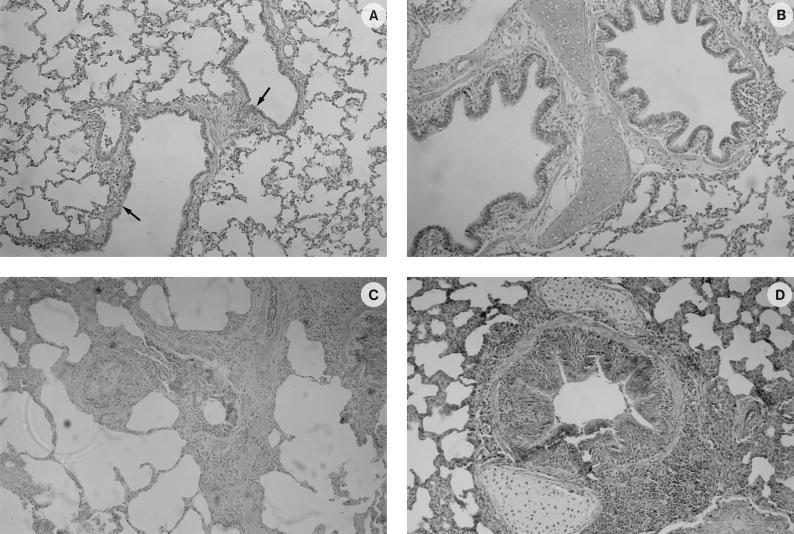
FIG. 7.
Histological findings in the lungs of control (A and B) and experimentally infected (C and D) SPF pigs as shown by HPS staining. (A) Spongiform aspect of the lung from a healthy pig, showing clear airway passages (bronchioli and alveolar duct indicated by arrows) and well-delineated interalveolar septae. (B) Normal appearance of a bronchiolus. (C) General aspect of the lung of a pig that has been infected with the IAF-DM9827 strain of M. hyopneumoniae, with characteristic perivascular and peribronchiolar mononuclear cells infiltration. No damage to the alveoli was observed, with only a mild mononuclear infiltration of the alveolar septae. (D) Peribronchiolar and perivascular accumulation of mononuclear cells with a mild hyperplasia of the bronchiolar epithelium.

