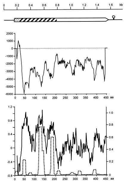
FIG. 1.
Structure of LppQ. (Top) Genetic structure of the 1,764-bp segment of M. mycoides subsp. mycoides SC strain Afadé, cloned in plasmid pJFFmaO5. The box represents the ORF of lppQ. Dotted segment, precursor signal sequence; hatched segment, antigenic, surface-exposed N-terminal half; open segment, integral membrane C-terminal half. The circle with a stem represents the transcriptional stop signal. (Middle) Transmembrane helix prediction diagram; aa, amino acids. (Bottom) The solid line and left-hand scale represent the hydrophilicity diagram calculated according to the method of Hopp and Woods (20); the dotted line and right-hand scale show the predicted value of the coiled-coil tertiary structure calculated using a window size of 14 aa on the Lupas scale (22).

