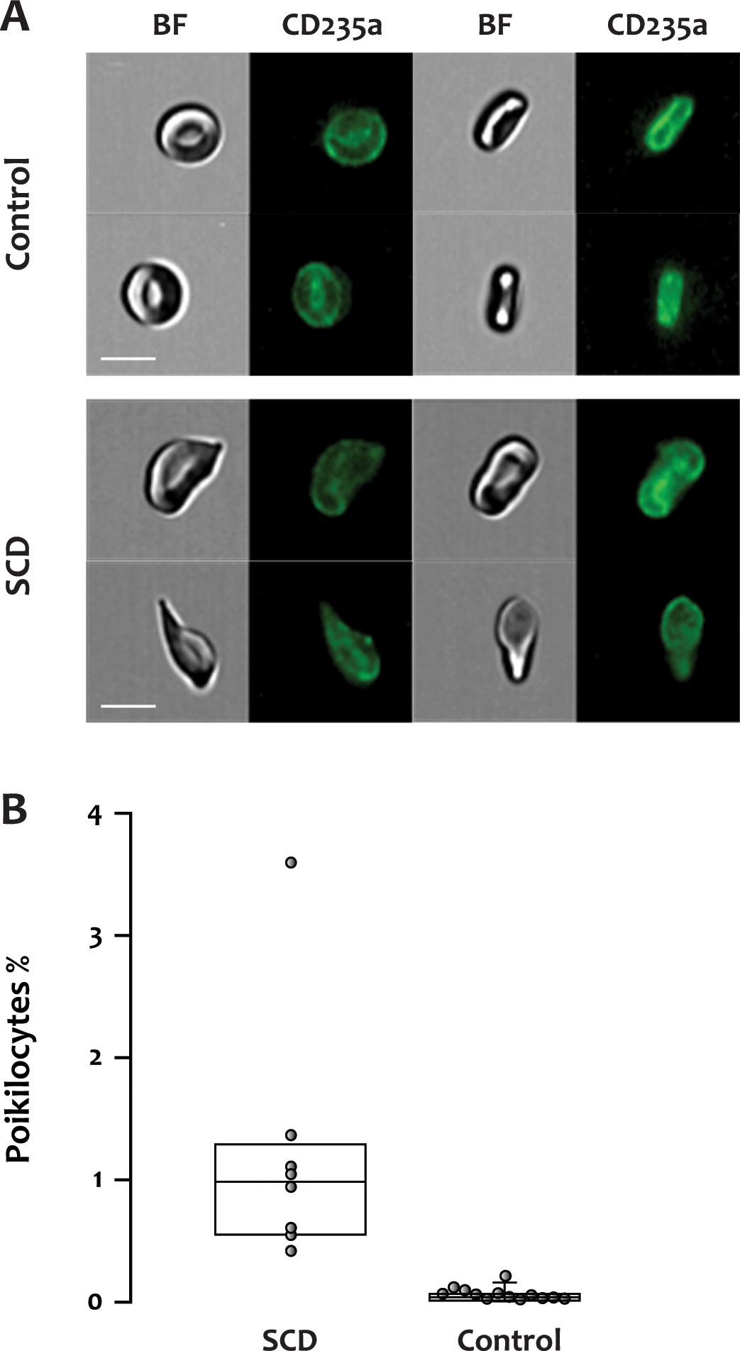
Figure 2. Poikilocytes in patients with SCD.

(A) Representative fluorescent and brightfield images of normally shaped RBCs from a healthy donor or poikilocytes from an SCD patient. BF: brightfield, CD235a: glycophorin A, scale bar: 7 μm. (B) The percentage of poikilocytes was significantly higher in the SCD patients (median, IQR: 0.984%, 0.567–1.224%, 14 samples from 8 patients) than in healthy donors (0.04%, 0.015–0.055%, p<0.001, 13 samples from 13 donors).
