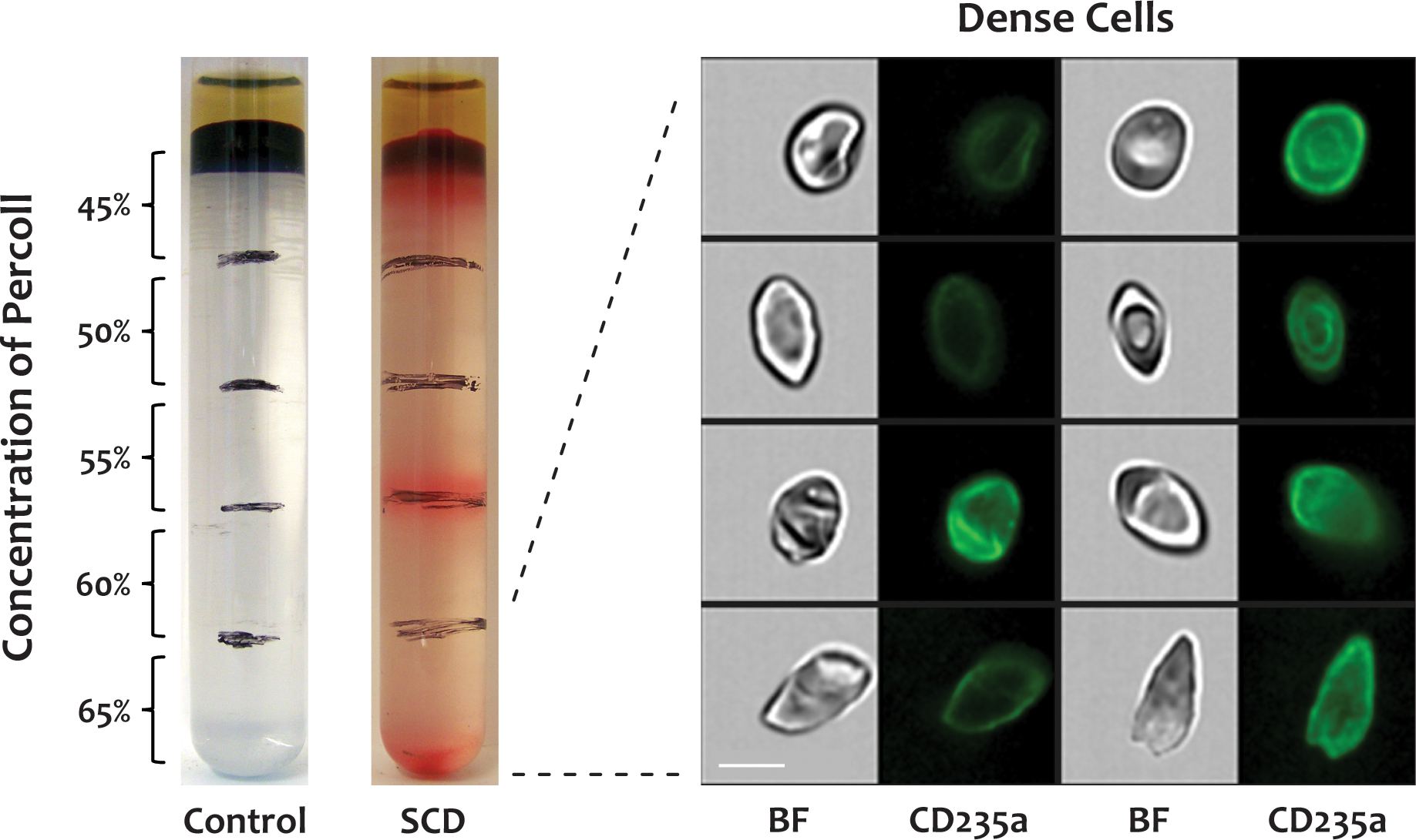
Figure 3. Dense cell morphology.

Dense cells were isolated by centrifuging whole blood from SCD patients through a discontinuous Percoll density gradient, labeled with a FITC-conjugated antibody to CD235a, and analyzed by IFC. The dense cells in the bottom fraction (65%) of the Percoll density gradient displayed diverse morphologies, similar to poikilocytes in whole blood. There were no dense cells in blood from healthy donors. BF: brightfield, CD235a: glycophorin A, scale bar: 7 μm.
