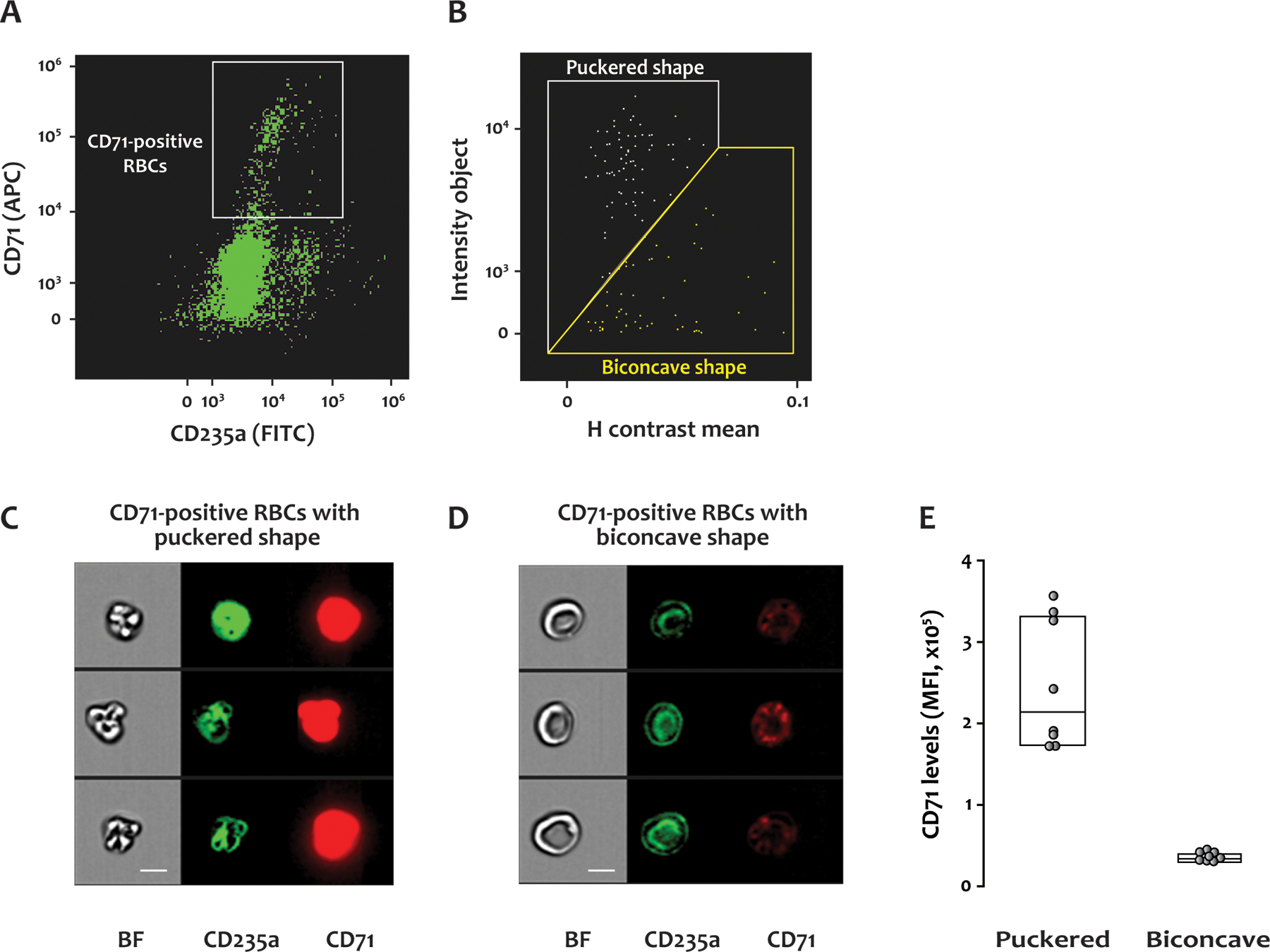
Figure 4. Morphologies of CD71-positive RBCs in SCD blood.

Citrated whole blood from SCD patients was labeled with antibodies to CD235a (FITC) and CD71 (APC), fixed with paraformaldehyde, and analyzed by IFC. (A) A scatter plot of RBCs with a gate for those positive for CD71. (B) CD71-positive RBCs with puckered shape were separated from those with biconcave shape using the plot of H contrast mean vs intensity object. (C) and (D) show representative brightfield and fluorescent images of the CD71-positive RBCs with puckered and biconcave morphologies, respectively. BF: brightfield, CD235a: glycophorin A, CD71: transferrin receptor, scale bar: 7 μm. (E) The level of CD71 was higher on the cells with puckered morphology than those with biconcave shape. MFI: Mean fluorescence intensity. Fourteen samples from 8 SCD patients were examined by IFC.
