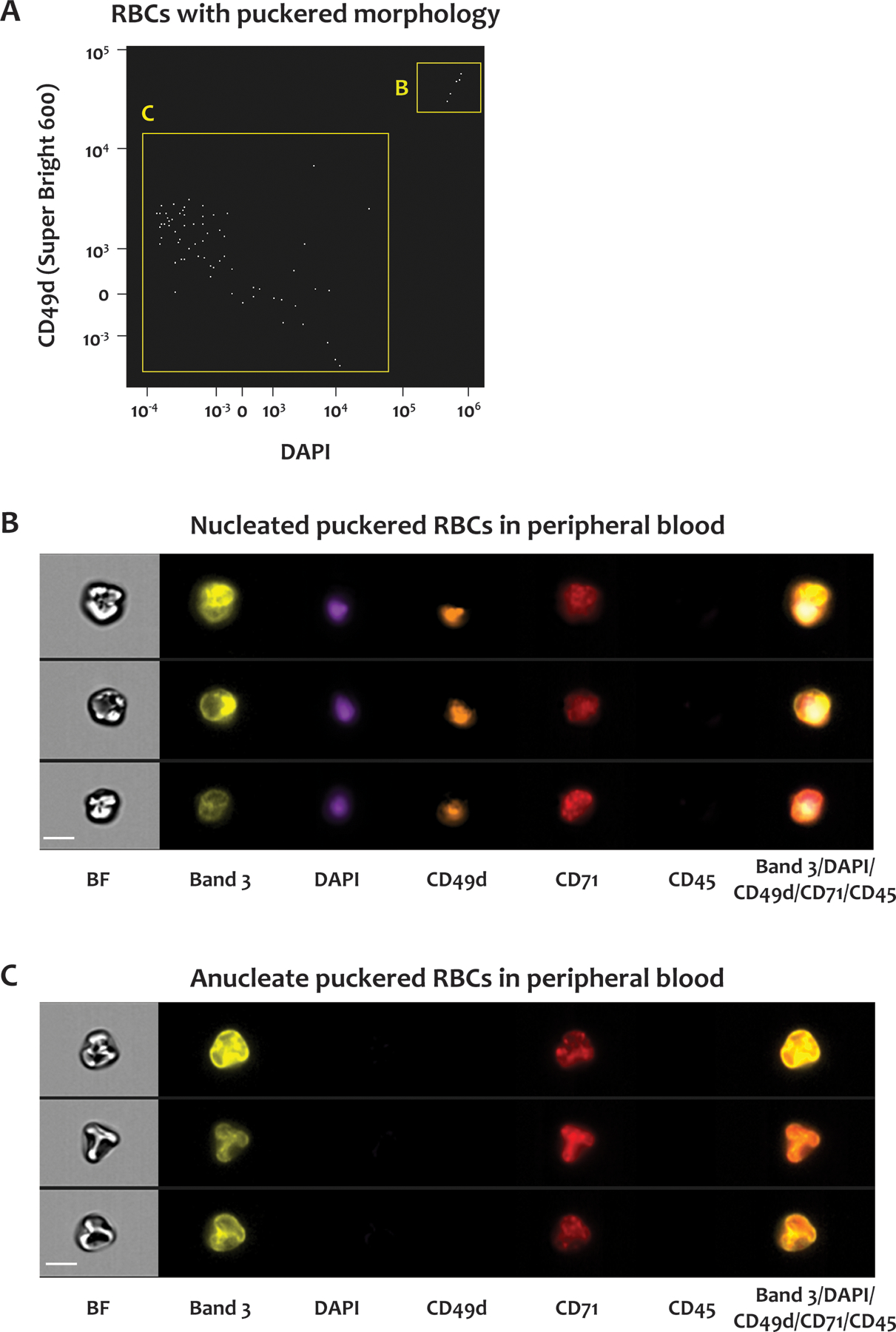
Figure 5. Nucleated RBCs in SCD blood.

Whole blood from SCD patients was stained for CD45, CD71, DNA, CD233 and CD49d. (A) Scatter plot of DAPI signals versus CD49d signals of CD71-positive RBCs with puckered morphologies. The RBCs in the B and C gates were positive and negative for both DAPI and CD49d, respectively. Representative brightfield and fluorescent images of the RBCs in the B and C gates in (A) are shown in panels (B) and (C), respectively. BF: brightfield, CD233: band 3, CD49d: α–4 integrin, CD71: transferrin receptor, CD45: leukocyte common antigen. Scale bar: 7 μm.
