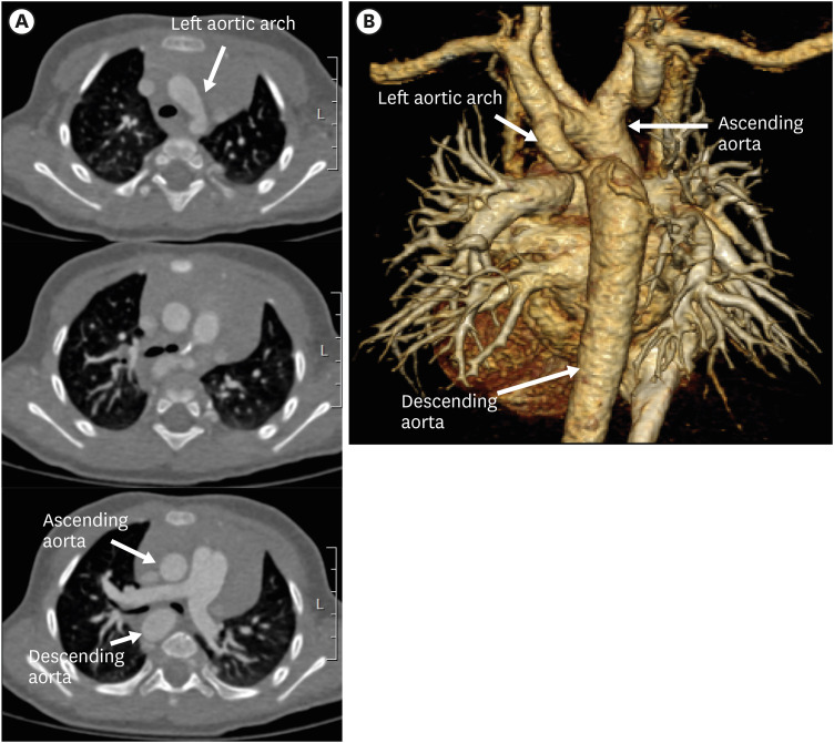
Figure 20. Left circumflex aorta in a 3-year-old boy. (A) Serial axial CT angiographic images and (B) 3D volume-rendered CT angiographic image show a left aortic arch crossing the midline posterior to the trachea and further descending on the right side of spine. Additionally, aortic arch hypoplasia and coarctation of the left aortic arch are present.
CT: computed tomography.

