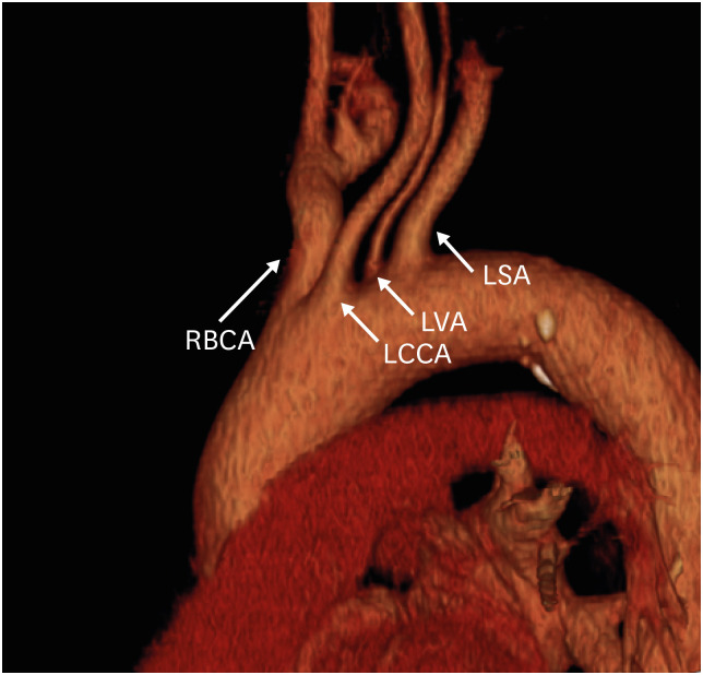
Figure 4. Direct origin of the left vertebral artery from the aortic arch in a 53-year-old man. Oblique sagittal 3D volume-rendered computed tomography angiographic image shows the direct origin of the LVA from the left aortic arch.
LCCA: left common carotid artery, LSA: left subclavian artery, LVA: left vertebral artery, RBCA: right brachiocephalic artery.

