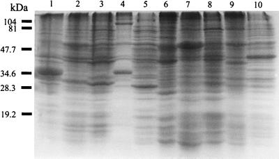
FIG. 1.
Coomassie brilliant blue staining profiles of Bartonella strains after SDS-PAGE. Lanes: 1, B. bacilliformis; 2, B. henselae serotype Houston; 3, B. henselae serotype Marseille; 4, B. clarridgeiae; 5, B. quintana; 6, B. elizabethae; 7, B. grahamii; 8, B. taylorii; 9, B. doshiae; 10, B. vinsonii. The positions of molecular mass markers of 104, 81, 47.7, 34.6, 28.3, and 19.2 kDa are noted on the left.

