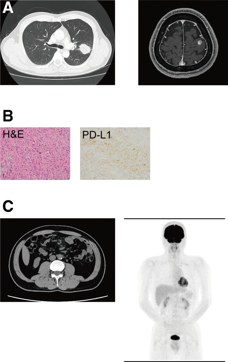
Figure 1.
An enlarging left lung mass (chest CT; left) and metastatic brain tumor (magnetic resonance imaging; right) at initial diagnosis (A). Pathological analyses of the resected tumor: hematoxylin and eosin (H&E) staining and immunohistochemical analysis for PD-L1 (B). Recurrence of pulmonary pleomorphic carcinoma was not detected by CT (left) or PET-CT (right) performed 3 weeks or 1 year, respectively, before this admission (C). CT = computed tomography, PD-L1 = programmed death-ligand 1, PET-CT = positron emission tomography-computed tomography.

