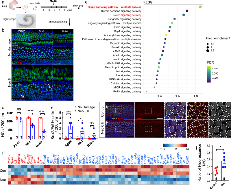
Fig. 1. Hippo signaling is involved during post-injury and self-repair processes in damaged neonatal mammalian cochleae in vitro.
a Cochleae of P2 neonatal mice were isolated and cultured for 12 h, then damaged with 1 mM neomycin for 6 h and cultured for another 5 days with 10 μM EdU. b, c After neomycin damage, HC death increased from the apex to the base of the cochlea (Myosin7a+ HCs, apex: 109.0 ± 6.4; middle: 76.7 ± 6.7; base: 43.0 ± 4.6), and two-way ANOVA followed by Sidak’s multiple comparison test showed that there was a significant difference in the middle and basal turns. d Very few proliferative SCs were observed after HC damage (Sox2/EdU double-positive SCs, apex: 2.4 ± 0.6; middle: 1.4 ± 0.5; base: 0.6 ± 0.4), although two-way ANOVA followed by Sidak’s multiple comparison test showed the significant difference in the apical and middle turns. e Kyoto Encyclopedia of Genes and Genomes (KEGG) pathway analysis of the transcriptome data. f Heatmap showing relative expression levels of the differentially expressed genes associated with the Hippo signaling pathway after HC damage. (FDR < 0.01; n = 3 for each condition). Higher expressed genes are shown in red, while the genes with relatively lower levels are depicted in blue. g–i Cochleae of P2 mice were dissected and cultured for 12 h and then treated with 1 mM neomycin for 6 h. Immunohistochemical staining was conducted after another 24 h culture. Compared with control groups, immunohistochemical staining and relative fluorescence quantitative analysis results analyzed with two-tailed, unpaired Student’s t tests showed that YAP (red) nuclear accumulation in Sox2+ SCs (white) was increased after neomycin injury. *p < 0.05, **p < 0.01, ***p < 0.001, ****p < 0.0001. Data are shown as the mean ± SEM. Scale bars = 20 μm.

