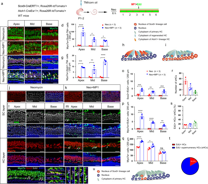
Fig. 5. YAP nuclear translocation enhances HC regeneration after neomycin damage in vitro.
a Sox9-CreERT2/+; Rosa26R-tdTomato/+ and Atoh1-CreEsr1/+; Rosa26R-tdTomato/+ mouse models were used to trace the SC and HC lineages. Tamoxifen was injected at P1 to activate tdTomato expression overnight, and the cochleae were harvested at P2 and then cultured in vitro. After neomycin exposure for 6 h, the cochlear explants were cultured with exogenous XMU-MP-1 or 0.5% DMSO for 5 days. b–d Compared with controls, the numbers of Myosin7a+/Sox9-tdTomato+ HCs were largely increased from the apical to basal turn in the XMU-MP-1-treated group analyzed with two-way ANOVA followed by Sidak’s multiple comparison test. e–g There were large numbers of Myosin7a+/Atoh1-tdTomato– HCs in the XMU-MP-1-treated groups compared to controls analyzed with two-way ANOVA followed by Sidak’s multiple comparison test. h The schematic diagram of Myosin7a+/Sox9-tdTomato+ HCs after XMU-MP-1 treatment. i The schematic diagram of Myosin7a+/Atoh1-tdTomato– HCs after XMU-MP-1 treatment. j, k The cochlear explants from P2 WT mice were harvested and then cultured. After neomycin exposure for 6 h, the explants were cultured with exogenous XMU-MP-1 or 0.5% DMSO for 24 h. Immunohistochemical staining results showed that the addition of XMU-MP-1 significantly increased the nuclear accumulation of YAP1 in Sox2+ SCs after neomycin injury. l, m After neomycin injury, the cochleae were cultured with DMSO or XMU-MP-1 for another 5 days, and 10 μM EdU was added the whole time to trace proliferating cells. In the control group with DMSO treatment, no proliferating HCs and very few SCs were detected. In the XMU-MP-1-treated group, there were some EdU+ SCs and HCs from the apical to basal turn. n The EdU+ HCs were mostly distributed in the tunnel between the inner and outer HCs, and they mostly appeared in pairs and stained with Sox2. o–q Compared with the control groups, there was a significant increase in the number of proliferating SCs and HCs after XMU-MP-1 addition in vitro analyzed with two-way ANOVA followed by Sidak’s multiple comparison test. The total number of HCs was significantly increased. r–t Only about 16% of HCs were EdU+ when compared with the increased proportion of HCs in the apical turn; sHC: supernumerary HC. u The schematic diagram of the mitotically regenerated HCs and proliferating SCs. *p < 0.05, **p < 0.01, ***p < 0.001, ****p < 0.0001. Data are shown as mean ± SEM. Scale bars = 20 μm.

