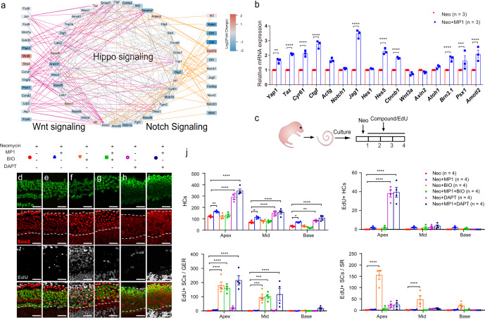
Fig. 7. Notch pathway inhibition works synergistically with Hippo-off to promote HC regeneration.
a Protein–protein interaction network analysis of differentially expressed genes of the Hippo, Notch, and Wnt signaling pathways. Purple lines indicate interactions between Hippo and Wnt genes, and orange lines indicate interactions between Hippo and Notch genes. The color of the nodes indicates the gene expression fold change. b The cochlear explants from P2 wild-type mice were harvested and then cultured. After neomycin exposure for 6 h, the explants were cultured with XMU-MP-1 or DMSO for 24 h. Related mRNA expression levels between XMU-MP-1 treated groups and controls are shown, and two-way ANOVA followed by Sidak’s multiple comparison test was performed to show the significant differences. c The cochlear explants from P2 WT mice were harvested and then cultured. After neomycin exposure for 6 h, the explants were cultured with exogenous compounds (including XMU-MP-1, BIO, or DAPT) or 0.5% DMSO for 3 days, and 10 μM EdU was added the whole time to trace proliferating cells. d–g The total numbers of HCs and EdU+ HCs were not significantly different between the XMU-MP-1-treated and XMU-MP-1/BIO-treated groups. e, h, i The number of regenerated HCs was significantly increased in the XMU-MP-1/DAPT-treated group when compared to the XMU-MP-1-treated group. j Two-way ANOVA followed by Dunnett’s multiple comparisons test was performed and showed that the upregulation of Wnt signaling by BIO could promote the proliferation of SCs. However, XMU-MP-1/DAPT co-regulation generated more HCs, including EdU+ HCs, and large numbers of proliferating SCs were found in the GER. *p < 0.05, **p < 0.01, ***p < 0.001, ****p < 0.0001. Data are shown as the mean ± SEM. Scale bars = 20 μm.

