Figure 3.
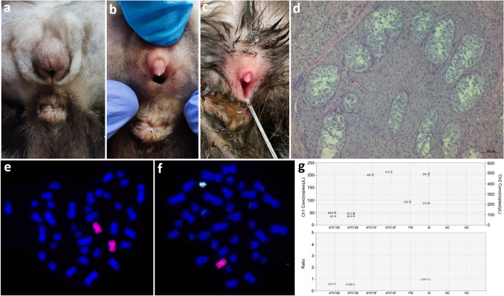
Characteristics of DSD case #7513: (a, b, c) ambiguous external genitalia: rudimentary penis and vulva; (d) histology of gonads: testes with sparse seminiferous tubules, hypertrophic Sertoli cells, numerous Leydig cells and lack of germinal epithelial cells; FISH with X (red) and Y (green) chromosome painting probes showing two cell lines: 38,XX (e) and 38,XY (f) in lymphocytes; (g) estimation of the Y/X ratio based on proportion of AMELY and AMELX genes in blood leukocytes (#7513B) and fibroblasts (#7513F). The upper chart shows amplification signals from chromosome X (green color) and Y (blue color). In fibroblasts of case #7513 (#7513F) the signal was detected from chromosome X only. Both signals were detected in blood leukocytes (#7513B). On the lower chart, an Y/X ratio of approximately 0.5 was detected in blood leukocytes (#7513B), indicating the presence of two cell lines. In fibroblasts of DSD case #7513 (#7513F) and in a control female (FM), the lack of Y chromosome resulted in a Y/X ratio of = 0, while a Y/X ratio of approximately 1 was detected in a control male (M); NC: negative control.

