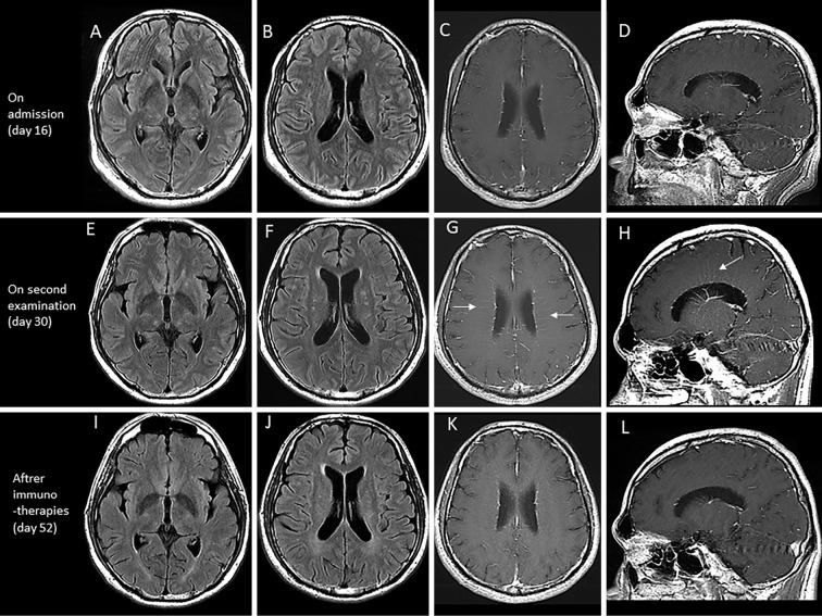
Figure 1.
Brain magnetic resource imaging (MRI) at 16 days (on admission) (A-D), 30 days (two weeks from admission) (E-H), and 52 days (after immunotherapies) (I-L) after the onset. Brain MRI on admission showed high intensity areas on fluid attenuated inversion recovery (FLAIR) imaging (A, B), but no evidence of periventricular gadolinium enhancement on T1 imaging (C, D). After two weeks, brain MRI showed increased high intensity areas on FLAIR imaging (E, F), and periventricular gadolinium enhancement (arrows) on T1 imaging (G, H). Brain MRI after immunotherapies showed that the spotty lesions had become paler (I, J), and there was a reduction in the enhancement (K, L).

