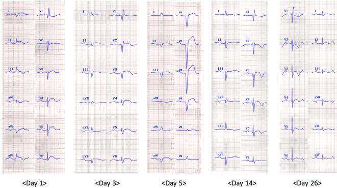
Figure 2.
Time-course changes of an electrocardiogram (ECG) after admission. The ECG on day 1 showing bifascicular block (first-degree atrioventricular block and complete right bundle branch block) and ST-segment elevation in leads I, II, III, aVF, V3, V4, V5 and V6 without reciprocal ST-segment depression. The ECG follow-up on day 3 showing trifascicular block (first-degree atrioventricular block, complete right bundle branch block, and left anterior hemiblock). The ECG on day 5 showing progression to atrioventricular dissociation. The ECG on day 14 showing the recovering trifascicular block, which transformed into a bifascicular block on day 26.

