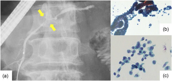
FIGURE 2.

Findings of stage 0 pancreatic ductal adenocarcinoma (Case 5 in Table 4, carcinoma in situ). (a) Endoscopic retrograde cholangiopancreatography. Pancreatography revealed stenosis of the main pancreatic duct in the body of the pancreas. The patient had no mass detected in other imaging studies. An endoscopic naso‐pancreatic drainage tube was placed, and serial pancreatic juice aspiration cytology was performed. (b, c) Images of liquid‐based cytology. The background of inflammatory cells and artifacts is removed, and solitary, scattered tumor cells can be evaluated.
