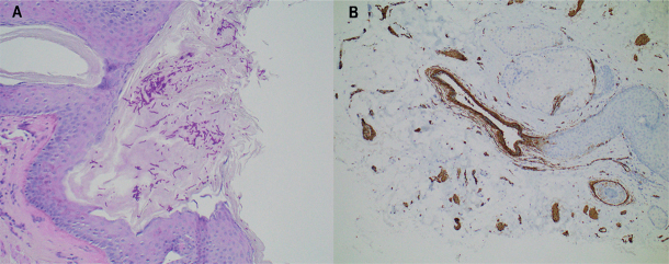
Fig. 1.
Histology. (A) Punch biopsy from a slightly elevated lesion on the perineum. Fungi, mainly in the form of hyphae, can be seen on the epithelium. The inflammatory response is sparse. The process in question is a superficial fungal infection. Periodic acid-Schiff (PAS) stain; original magnification ×200. (B) Punch biopsy from a plaque-like, elevated perineal lesion. Histologically smooth muscle fibres unrelated to the follicular apparatus are haphazardly spaced, unlike in the normal dermis. The findings are consistent with (acquired) smooth muscle hamartoma. Staining for smooth muscle actin (SMA) highlights the smooth muscle fibres (brown structures); original magnification ×100.

