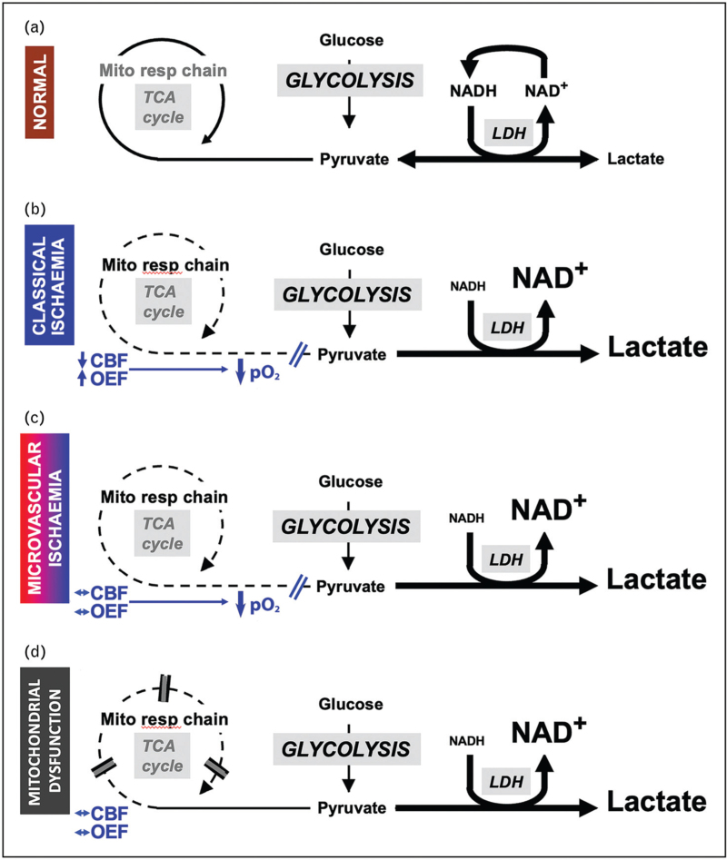
FIGURE 1.
Schematic to demonstrate derangements in glucose metabolism following traumatic brain injury. Cerebral blood flow (CBF), lactate dehydrogenase (LDH), mitochondrial respiratory chain (Mito resp chain), nicotinamide adenine dinucleotide and its reduced form (NAD and NADH), oxygen extraction fraction (OEF), partial pressure of oxygen (pO2), TCA (tricarboxylic acid cycle). Under normal conditions both glucose and lactate can be metabolized to pyruvate which enters the tricarboxylic acid cycle and undergoes oxidative phosphorylation. While the concentration of lactate is higher than that of pyruvate the lactate/pyruvate ratio remains less than 20 (a). In classical ischemia the increase in oxygen extraction fraction that results from a reduction in cerebral blood flow can no longer maintain oxygen delivery to the brain and tissue pO2 falls preventing mitochondrial oxidative phosphorylation. Pyruvate is converted to lactate via lactate dehydrogenase generating NAD which is required to maintain increased glycolysis and energy production, and the lactate/pyruvate ratio increases to levels more than 25 (b). A similar increase in glycolysis resulting in a lactate/pyruvate ratio more than 25 occurs in microvascular ischemia where microvascular thrombosis, collapse and perivascular edema result in an increased diffusion barrier that limits oxygen delivery leading to low tissue pO2 (c), and mitochondrial dysfunction where oxidative phosphorylation is suspended despite maintenance of normal oxygen and glucose delivery (d). In both (c) and (d), average macrovascular cerebral blood flow and oxygen extraction fraction values are not typically ischemic. Modified with permission from that originally published in Menon DK, Ercole A. Critical care management of traumatic brain injury. Handb Clin Neurol 2017;140:239–74.

