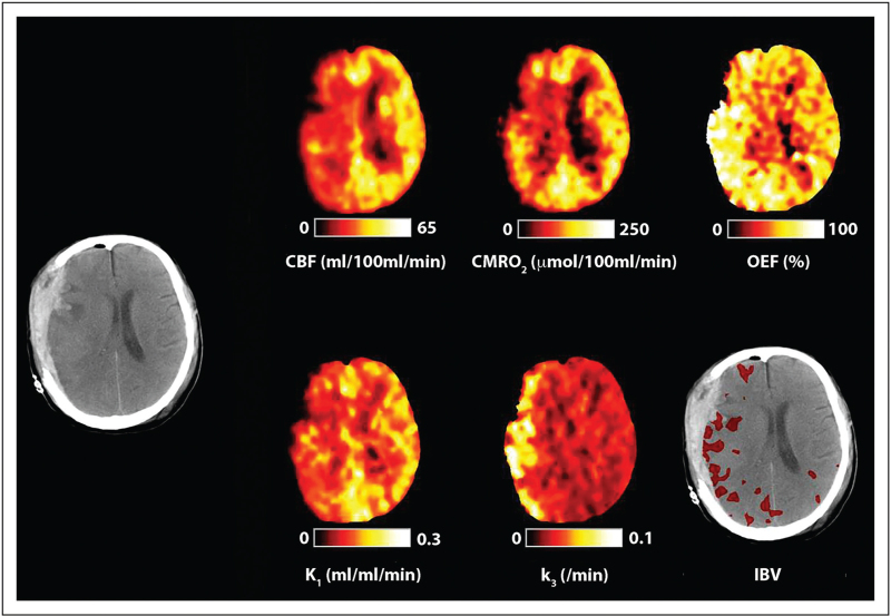
FIGURE 2.
Cerebral ischemia. Computed tomography, cerebral blood flow, cerebral metabolic rate of oxygen, oxygen extraction fraction, ischemic brain volume (IBV) and PET 18F-fluorodeoxyglucose kinetic parameters for K1 (glucose delivery) and k3 (glycolysis) obtained in a 40-year-old female within 24 h of severe traumatic brain injury resulting from a fall. The computed tomography scan was obtained following evacuation of a subdural hematoma and demonstrates residual subdural blood with minimal midline shift and underlying hemorrhagic contusions. Cerebral blood flow, cerebral metabolic rate of oxygen and (K1) glucose delivery are reduced, whereas oxygen extraction fraction and k3 (glycolysis) are increased within the hemisphere underlying the subdural. These findings are consistent with classical ischemia.

