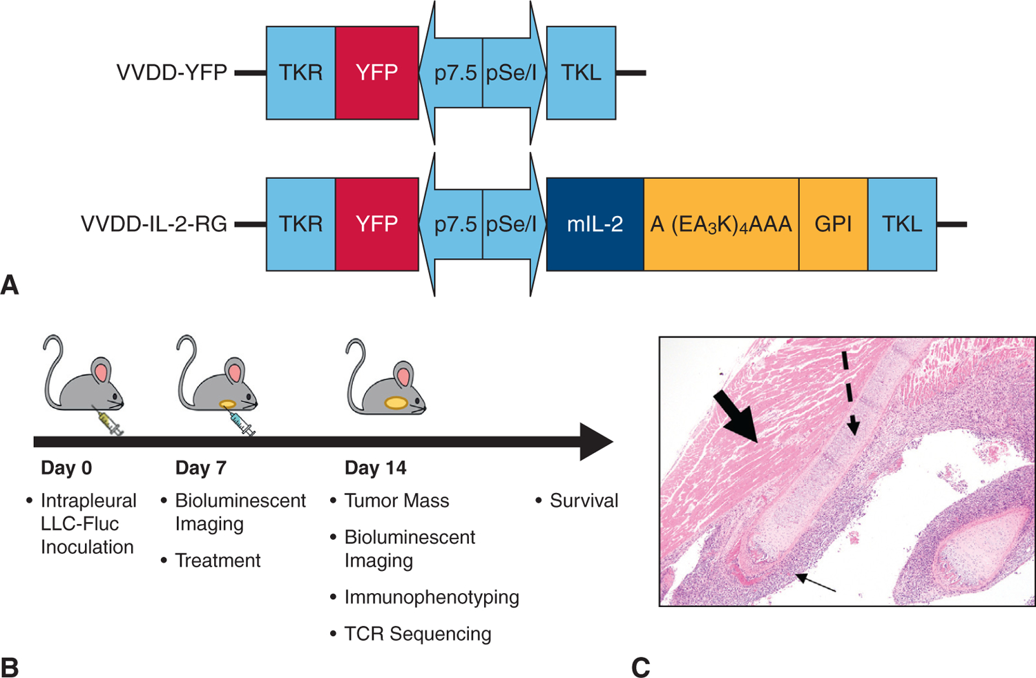
FIGURE 1.

VV variants and murine model of MPD methodology. A, Doubly deleted VV-YFP expressing membrane bound VV-IL-2 constructs. B, Illustrative display of the timing of experiments to assess day 14 end points and survival after inoculation with tumor and corresponding day 7 treatment. C, Hematoxylin–eosin stain of resected day 7 metastatic pleural disease (solid thin arrow), rib (dashed arrow), and intercostal muscle (bolded arrow) demonstrating successful tumor implantation in the MPD model (10× magnification).
