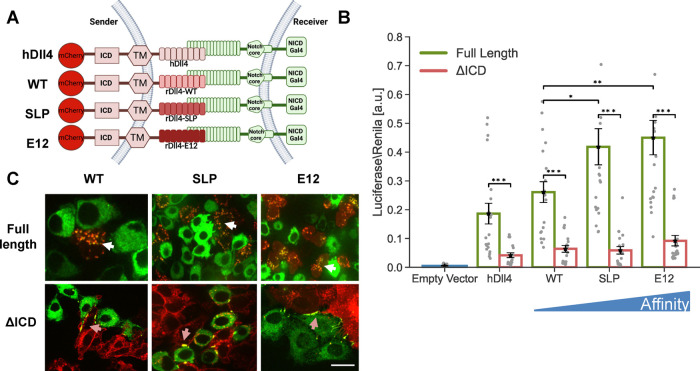
Figure 2.
Higher affinity receptor–ligand interactions do not compensate for the lack of ligand ICD. (A) Schematic of the chimeric variants of Dll4 generated for testing the role of receptor–ligand affinity. Here, hDll4 is the full-length human Dll4 ligand. All the chimeric variants contain human Dll4ICD and TM domains and different versions of rat Dll4ECD. Binding affinities of WT, SLP, and E12 to Notch1 are 12.7 μM, 440 nM, and 56 nM, respectively (Luca et al.21). All the variants are placed under an inducible promoter and are fused to mCherry. (B) Luciferase activity assay showing the activation of a Notch reporter cell line (U2OS-Notch1-Gal4 transfected with a UAS-Luciferase reporter) co-cultured with CHO-TetR cells expressing the indicated variants either with or without ligand ICD. (C) Images showing a co-culture of inducible affinity variants (red) with Notch1-citrine cells (green) 10 h after the induction of ligand expression with 100 ng/mL dox. TEC is observed in full-length ligands (white arrows). Accumulation on the boundaries, but no TEC, are observed in ligands lacking their ICD (orange arrows). Data points show mean values from n = 20 measurements for (B), from five independent experiments. Error bars represent S.E.M. *p < 0.05, **p < 0.01, ***p < 0.001. Scale bars-10 μm.

