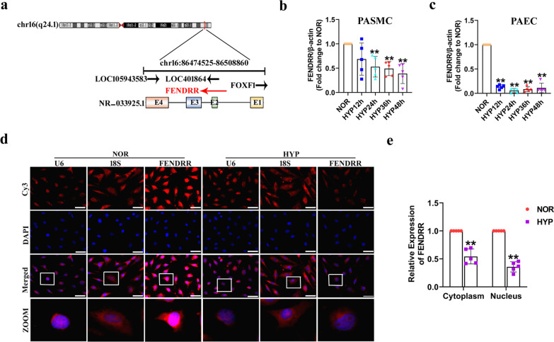
Fig. 1.
FENDRR expression is decreased under hypoxic conditions. a Genomic location of FENDRR. The arrows indicate the direction of transcription. b Expression of FENDRR quantified by qRT-PCR in human pulmonary artery smooth muscle cells endothelial cells (HPASMCs) (n = 5). c Expression of FENDRR quantified by qRT-PCR in human pulmonary artery endothelial cells (HPAECs) (n = 5). d HPAECs were cultured for 24 h under HYP conditions, and fluorescence in situ hybridization (FISH) assay was performed to detect FENDRR expression. U6 and 18S RNA were used as controls for localization of the nucleus and cytoplasm. Scale bar = 50 μm. e FENDRR expression in the nucleus and cytoplasm of HPAECs after exposure to HYP for 24 h (n = 5). Each datapoint in the figure represents a unique biological replicate. All values are presented as the mean ± SD. Statistical analysis was performed with Student’s t-test. NOR: normoxia; HYP: hypoxia. **P < 0.01 compared with NOR

