Fig. 5.
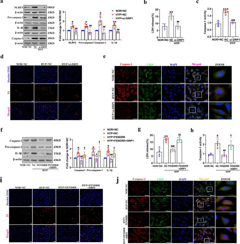
FENDRR regulates HPAECs pyroptosis via DRP1. a After transfection with DRP1 siRNA, Western blot was used to examine the protein levels of Caspase-1, NLRP3, Pro-caspase-1 and IL-1β (n = 5). b LDH release assays were used to determine the effects of DRP1 on HPAECs pyroptosis (n = 5). c The activity of caspase-1 was examined using caspase-1 activity assay kit (n = 5). d Images of fluorescence staining with PI (red) and Hoechst 33,342 (blue) were used to detect PI-positive stained cells. Scale bar = 50 μm. e Immunofluorescence analysis of Caspase-1 (red) and CD31 (green) expression. Scale bar = 50 μm. f After cotransfection with FENDRR and DRP1 overexpression plasmid, Western blotting was used to examine the protein levels of Caspase-1, NLRP3, Pro-caspase-1 and IL-1β (n = 5). g and h LDH release assays and activity of caspase-1 were used to determine the effects of cotransfection with FENDRR and DRP1 overexpression plasmid on HPAECs pyroptosis (n = 5). i and j PI staining and fluorescence intensity of Caspase-1 were used to determine the effects of cotransfection with FENDRR and DRP1 overexpression plasmid on HPAECs pyroptosis. Each datapoint in the figure represents a unique biological replicate. All values are presented as the mean ± SD. Statistical analysis was performed with one-way ANOVA. NOR: normoxic; HYP: hypoxic; NC: negative control. **P < 0.01, *P < 0.05 compared with NOR + NC. #p < 0.05, ##p < 0.01, ###p < 0.001 versus HYP + NC. $p < 0.05, $$p < 0.01 compared with HYP + FENDRR

