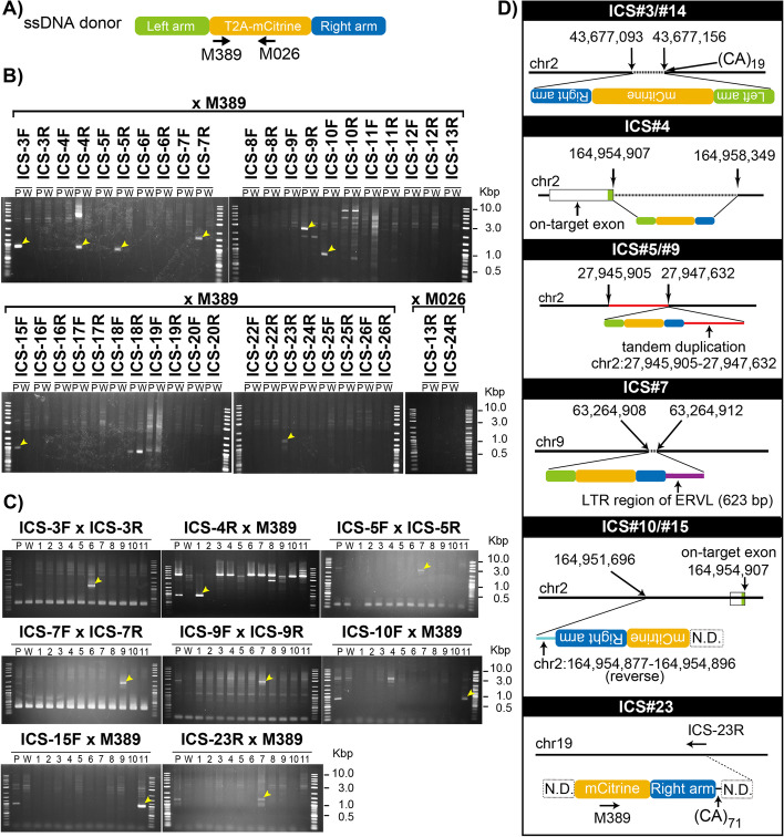
Fig. 4.
Detection and characterization of RIs and imprecise on-target insertions of the donor DNA in founder mice. A Location of PCR primers (M389 and M026) in the donor DNA cassette. B PCR screening of insertion sites for 22 ICSs using the pooled DNA derived from founder mice (P) and wild type (W) as templates. Yellow arrows indicate the fragments amplified only from founder mice. C PCR screening of the individual founder mice for eight ICSs detected by PCR in (B). Yellow arrows indicate the fragments uniquely amplified from founder mice. D Schematic for configuration of the inserted sequence. The junctional sequences of six loci (from nine ICSs) were analyzed by Sanger sequence. Chromosome positions were obtained from the UCSC Mouse Genome Browser, mm10 assembly. (CA)19 and (CA)71 indicate the length of 19 and 71 CA repeats, respectively. Dashed horizontal lines indicate deleted region of the genome. The solid red line indicates the tandemly duplicated region with mCitrine cassette. The purple line indicates a long terminal repeat (LTR) region of the endogenous retrovirus (ERVL). N.D., not determined

