
Figure 3. Visual cortex and superior colliculus are not required for contagious itch.
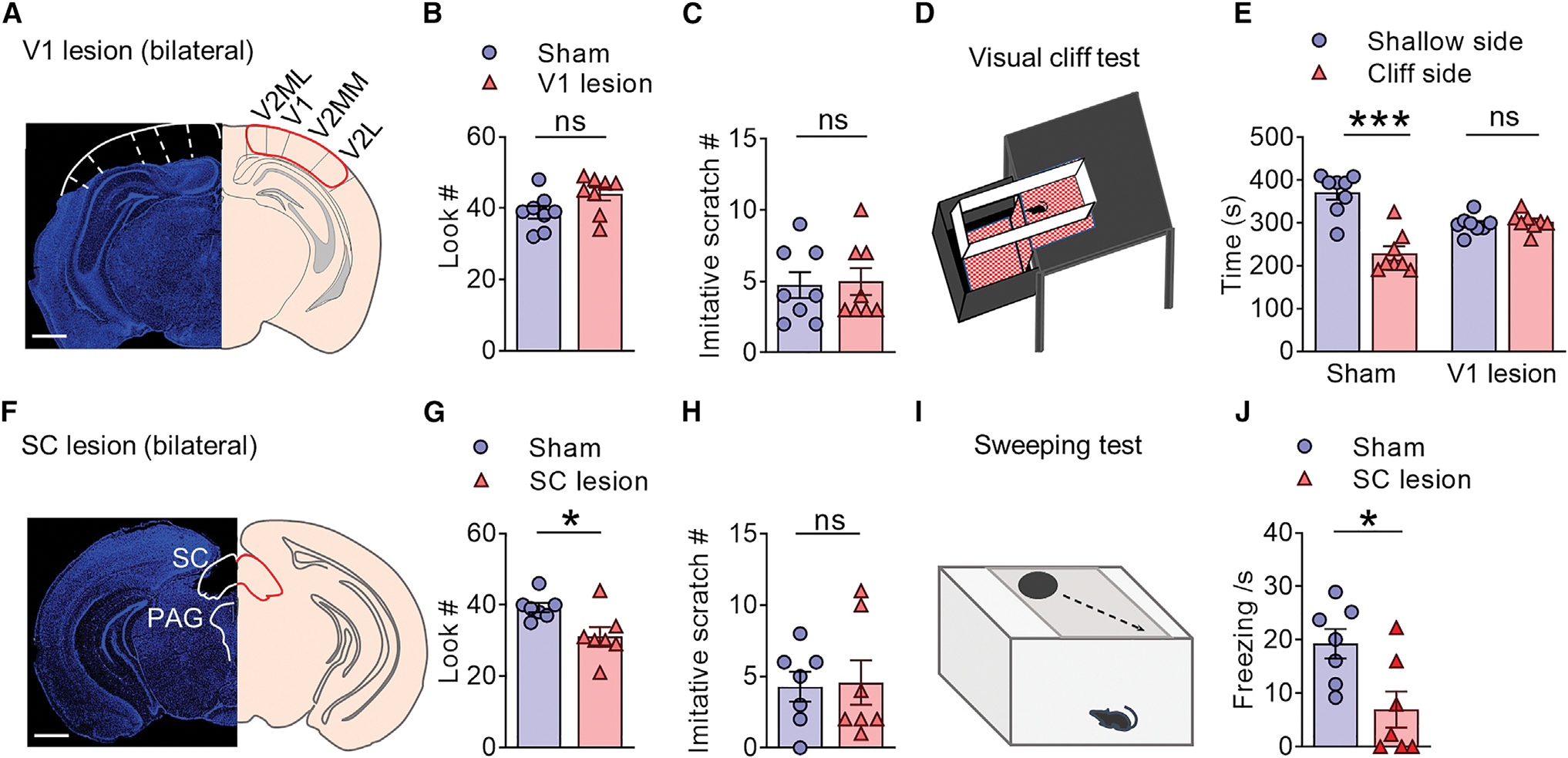
(A) Coronal section of the brain stained with DAPI (blue) showing visual cortex lesion (left) and corresponding anatomical location (right). Scale bar, 500 μm.
(B and C) Mean number of look (B) (t = 1.986, df = 14, p = 0.0670) and imitative scratch behaviors (C) (t = 0.1895, df = 14, p = 0.8524) of mice with visual sham surgery and visual cortex lesion.
(D) Cartoon illustrating the visual cliff test.
(E) Left: mice with sham surgery spent significantly shorter time on the cliff side than the shallow side, indicating a normal depth perception. Right: mice with visual cortex lesion spent a comparable amount of time on either side, indicating a loss of depth perception. F(1, 14) = 31.94, p < 0.0001.
(F) Coronal section of the brain (blue, DAPI) showing SC lesion (left) and corresponding anatomical location. SC, superior colliculus. PAG, periaqueductal gray. Scale bar, 500 μm.
(G and H) Mean number of look (G) (t = 2.752, df = 12, p = 0.0175) and imitative scratch behaviors (H) (t = 0.1516, df = 12, p = 0.8820) of mice with SC lesion.
(I) Cartoon illustrating the sweeping test.
(J) SC lesion significantly reduces freezing behavior during sweeping test relative to the control (t = 2.818, df = 12, p = 0.0155). n = 8 mice/group for V1 lesion, n = 7 mice/group for SC lesion.
Data are presented as mean ± SEM. Unpaired t test in (B, C, G, H, and J). Two-way ANOVA in (E). ns, not significant. *p < 0.05, ***p < 0.001.