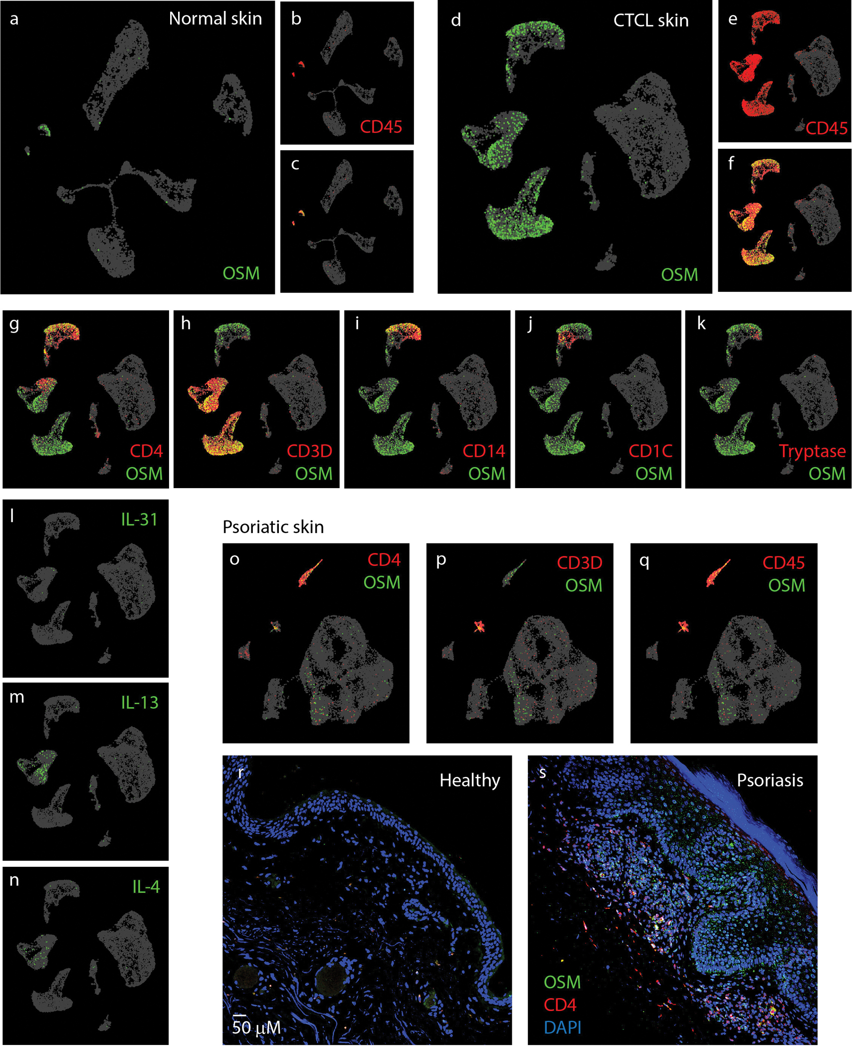
Figure 4. OSM is predominantly expressed in skin T-cells.

(A-C) UMAP plot of single-cell RNA-sequencing data (GSE128531) pooled from 4 normal skin biopsies (14,179 cells) showing expression pattern of OSM (green) and pan-leukocyte marker CD45 (PTPRC) (red). (D-F) UMAP plot of GSE128531 dataset which included 5 CTCL skin biopsies (30,663 cells) showing expression of OSM (green) and CD45 (red). (G-K) Co-expression of OSM (green) with markers for immune cell genes CD4+ and CD3+ (T-cells) (G-H), CD14+ (monocytes) (I), CD1C (dendritic cells) (J), and tryptase (mast cell) (K). (L-N) Expression of IL-31, IL-13, and IL-4 as indicated. (O-T) UMAP plots of single-cell sequencing data (EGAS00001002927) of psoriatic skin biopsies (21,025 cells pooled from 3 patients). Co-expression of OSM with CD4 (O), CD3 (P) and CD45 (Q). (R-S) Representative images of parafilm-embedded human skin sections immuostained against OSM (green) and CD4 (red) in healthy controls(R) and in psoriatic skin samples (S)
