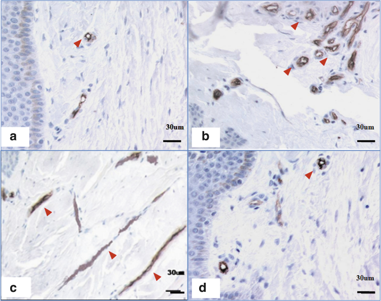
Figure 2.
CD34 staining of microvessel density in scars of different stages. (a) A few microvessels were apparent in normal skin. (b) Many microvessels were apparent in a proliferative scar. (c) Most microvessels were occluded in an RS. (d) The number of microvessels in a mature scar was comparable to that in normal skin. Scale bar = 30 μm. The red arrows indicate the microvessel. RS, regressive scar. Color images are available online.

