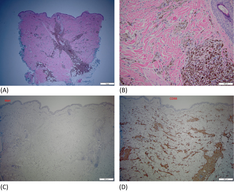
Figure 2. Pathology.
(A) H&E-stained section showing a heavy infiltrate of pigmented cells in the dermis in perivascular and perifollicular distribution, magnification 2x. (B) Their bland cytology and coarse melanin granules, magnification 20x. (C) Immunostain for Mart (melanoma marker) is negative, magnification 4x (note: endogenous melanin pigment has been removed for immunostain study). (D) Immunostain for CD68 (histiocyte marker) is diffusely positive in the same area, confirming that pigmented cells are melanophages (melanin-laded macrophages), magnification 4x.

