Figure 5.
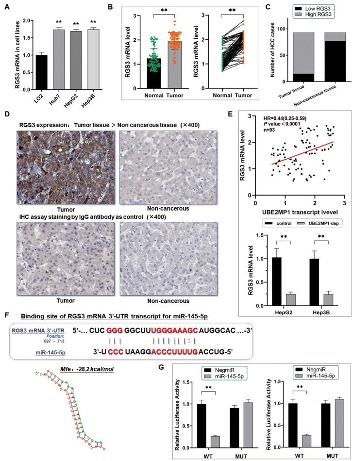
RGS3 mRNA is targeted by miR-145-5p post-transcriptionally. (A) The RT-qPCR assay demonstrated a significant up-regulation of RGS3 mRNA in three HCC cell lines, in comparison with the control LO2 cells (**P<0.01). (B) RT-qPCR assay was conducted on 93 real patients’ specimens. The RGS3 was significantly highly expressed in tumor tissues (**P<0.01). (C) Statistic of the number of cases concerning the expression of RGS3 in HCC specimens. RGS3 is highly expressed in most of the tumor tissues (78/93) (P<0.01). (D) Representative graph of immunohistochemistry analysis (400×) of the HCC cases. The IgG antibody was used for staining the specimens as a control. RGS3 expression in tumor specimens was significantly higher than in adjacent non-cancerous tissues. (E) RGS3 shared a positively correlated with UBE2MP1 expression in the HCC tissues from our center, and the expression of RGS3 presented a remarkable decrease in HCC cells when UBE2MP1 was depleted (**P<0.01). (F) Predicted binding sequence of the 3’UTR of RGS3 mRNA with miR-145-5p. The Mfe value is calculated as: -28.2 kcal/mol. (G) The dual-luciferase reporter assay verified the direct interaction between miR-145-5p and RGS3 mRNA (**P<0.01).

