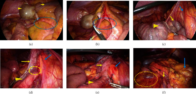
Figure 3.

The operative findings in Case 1. (a) The small intestine run through the hernia defect (dotted area), which consists of the fibrotic adhesions between the retroperitoneum and the colon mesentery. Cecum (arrowhead) and terminal ileum (arrow) were recognized over the hernia gate. (b) The hernia defect (dotted area) was dissected. (c) The outside adhesion of the right colon and the abnormal retroperitoneal bands was also dissected. After that, the duodenum (yellow arrow) and the jejunum (yellow arrowhead) were recognized under the ascending colon (blue arrow). (d) There was a severe curve (dotted area) at the transition from the proximal side of the duodenum (yellow arrow) to the distal side (yellow arrowhead), and a membrane structure ran across the duodenum and the pancreas head (blue arrow). (e) The transition between the proximal side of the duodenum (yellow arrow) and the distal side (yellow arrowhead) was straightened. The bleeding was recognized at the pancreatic head (blue arrow). (f) The small intestinal mesentery (dotted area) was recognized at the right side, and the transverse colon (blue arrow) was at the left side. The root of the small intestinal mesentery (yellow arrow) was fully widened.
