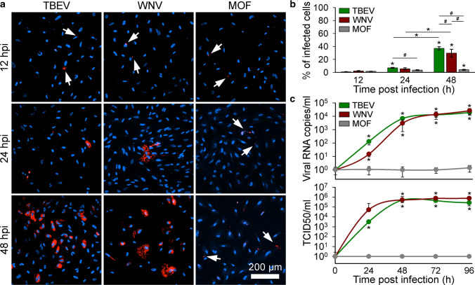
Fig. 4.
Tick-borne encephalitis virus (TBEV), West Nile virus (WNV), and mosquito-only flavivirus (MOF) infect human astrocytes, but only TBEV and WNV replicate efficiently in human astrocytes. a Human astrocytes infected with TBEV, WNV, and MOF and immuno-labeled with antibodies against flavivirus group antigen (red) at 12, 24, and 48 h post infection (hpi). Cell nuclei are stained with 4′,6-diamidino-2-phenylindole (DAPI; blue). Arrows indicate weakly infected cells. b The percentages of TBEV-, WNV- and MOF-infected cells at 12, 24, and 48 hpi were determined as the ratio between the number of flavivirus group antigen-positive cells and the number of DAPI-stained nuclei. Infection rates in human astrocytes increase with time for all three flaviviruses (*P < 0.05, one-way ANOVA followed by Dunn’s test). TBEV infection yields the highest percentage of infected cells at 48 hpi compared with WNV and MOF (#P < 0.05, one-way ANOVA followed by Dunn’s test). Data are presented as an average ± standard error. Statistical comparison was made between the groups of cells infected with the same virus at different hpi (*P < 0.05, one-way ANOVA followed by Dunn’s test) and between samples infected with different viruses at the same infection duration (#P < 0.05, one-way ANOVA followed by Dunn’s test). Number of cells analyzed at 12, 24, and 48 hpi: TBEV (1363, 1169 and 1550), WNV (674, 1047 and 1081), MOF (1010, 1064 and 773). Number of fields of view analyzed at 12, 24, and 48 hpi: TBEV (20, 20 and 20), WNV (19, 21 and 24), MOF (22, 22 and 21). c Replication rates of TBEV, WNV, and MOF in human astrocytes, determined by quantifying the amount of viral RNA in the cell culture supernatant (Viral RNA copies/ml; top graph) and the median tissue culture infectious dose (TCID50/ml; bottom graph) at different hpi (from 0 to 96). The concentration of TBEV and WNV RNA increases with time post infection, reaching the plateau at 48 hpi for both viruses (*P < 0.05, Mann–Whitney U test), whereas the quantity of MOF RNA does not change with time post infection in human astrocytes (P > 0.05, Mann–Whitney U test). TCID50 of TBEV and WNV confirms infectivity of astrocyte-released virus in Vero E6 cells and the concentration of infectious TBEV and WNV increases with time post infection (*P < 0.05, Mann–Whitney U test), while released MOF is not infective (P > 0.05, Mann–Whitney U test). Data are presented on a logarithmic scale as an average ± standard error. Statistical comparison was made against initial values obtained at 0 time point. Cells were infected with TBEV and MOF at an MOI 0.1 and with WNV at an MOI 1

