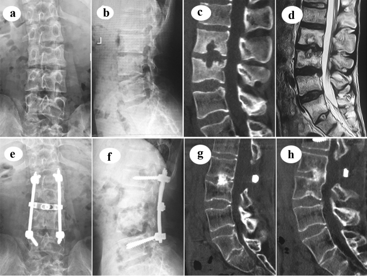
Fig. 3.
A 29-year-old male patient with L2–3 mono-segmental spinal TB, who complained of severe low back pain for 3 months, received single posterior debridement, interbody fusion and pedicle fixation. a, b Preoperative X-ray of AP and lateral images show roughly normal radiological presentation. c, d Preoperative CT and MRI images show the destruction of vertebral bodies and disc. e, f Postoperative X-ray of AP and lateral images show good position of pedicle screw fixation. g, h Postoperative CT images show solid interbody fusion had been achieved at the postoperative 24th month

