Figure 2.

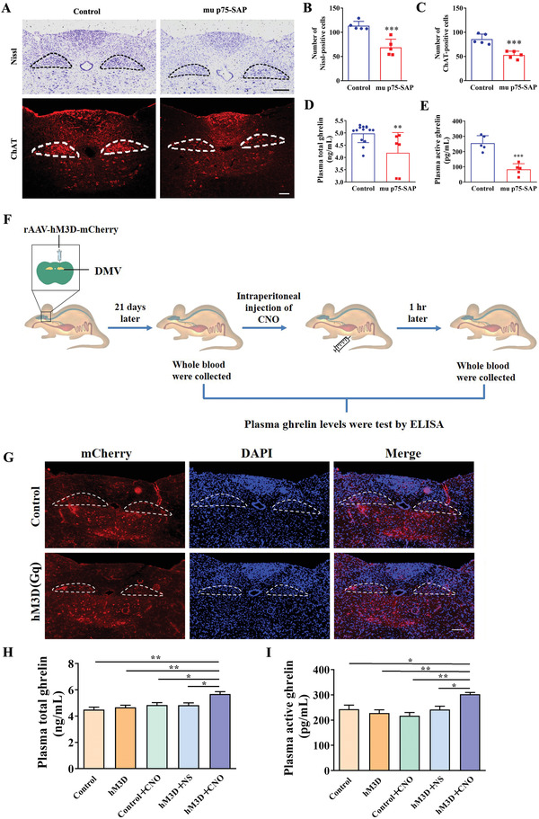
Changed plasma ghrelin levels depended on the activation of cholinergic neurons in the DMV. A) Representative images of Nissl‐positive cells and ChAT‐positive cells that stained by ChAT antibody of mice both in control and mu p75‐SAP‐treated group. Encircled areas: DMV region. Scale bar: 100 µm. B) Statistical analysis of Nissl‐positive cells. C) Statistical analysis of ChAT‐positive cells. D) Total plasma ghrelin levels were decreased in mu p75‐SAP‐treated mice group. E) Plasma active ghrelin levels were decreased in mu p75‐SAP‐amdinistered animals group. F) Schematic diagram of chemical genetic experiments. To assess if cholinergic DMV neurons are responsible for the secretion of ghrelin, cholinergic neurons in the DMV were infected by mCherry‐tagged hM3D (Gq) virus, which could be activated by CNO. The plasma ghrelin levels were analyzed before and after CNO injection. G) Expression of mCherry was observed in DMV region of ChAT‐cre mice with mCherry‐tagged virus injection. Encircled areas: DMV region. Scale bar: 100 µm. H) Effects of activation of cholinergic neurons in DMV on total plasma ghrelin levels. I) Effects of activation of cholinergic neurons in the DMV on active plasma ghrelin levels. B–E) Data were mean ± SEM (t‐test, **P < 0.01, ***P < 0.001, compared with the control), n ≥ 5 for each group. H,I) Data were mean ± SEM (One‐way ANOVA, followed by the Tukey test, *P < 0.05, **P < 0.01), n ≥ 5 for each group.
