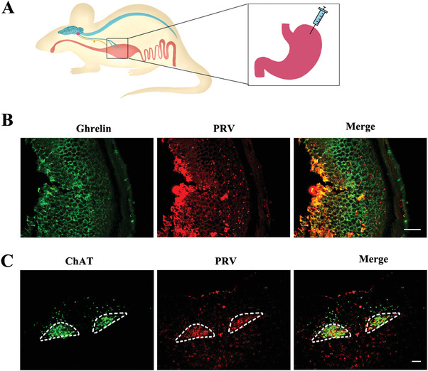
Figure 4.

Retrograde tracing of nerve fibers from the gastric fundus to the DMV region. A) Model of injection of PRV‐CAG‐EGFP virus in the gastric fundus. B) Co‐localization of PRV‐CAG‐EGFP expression and was observed on the ghrelin‐positive cells (stained by ghrelin antibody). Scale bar: 100 µm, n = 5. C) GFP was observed on the ChAT‐positive neurons that stained by ChAT antibody in the DMV area. Encircled areas: DMV region. Scale bar: 100 µm, n = 5.
