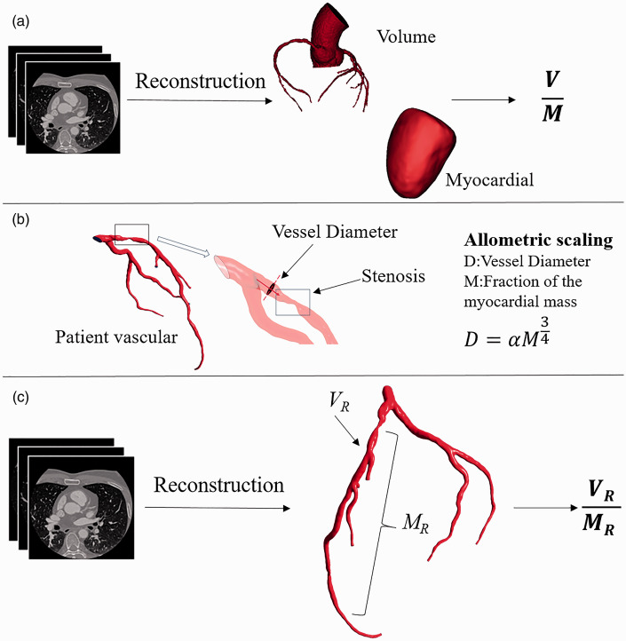
Figure 2.
Methodology for computing V/M and VR/MR. (a) The epicardial coronary arteries were segmented from coronary CTA data, and a three-dimensional volumetric model was created. The left ventricle myocardial volume was extracted from the CTA data and multiplied by a constant density value to obtain the myocardial mass. V/M is the ratio of epicardial coronary arterial lumen volume to left ventricle myocardial mass. 11 (b) Determination of the fraction of the myocardial mass by allometric scaling. 22 (c) Stenosis in the epicardial coronary arteries was segmented from coronary CTA data and the three-dimensional volumetric model. The fraction of the myocardial mass subtended by the vessel territory was calculated by allometric scaling. VR/MR is the ratio of regional coronary artery stenosis lumen volume to the fraction of the myocardial mass subtended by the vessel territory ratio. (A color version of this figure is available in the online journal.)

